A Novel High Definition Imaging (HDI) Informatics Platform
Dies ist ein Applikationsbericht, der keinen detaillierten Abschnitt zu Versuchen enthält.
Abstract
A brief overview of the new Waters® High Definition Imaging (HDI™) Software solution that allows the maximum information to be obtained from imaging experiments that combine ion mobility with mass spectrometry.
Introduction
Imaging using mass spectrometry is a rapidly expanding area that has extensively used MALDI ionization. Waters has pioneered the use of ion mobility spectrometry with MALDI imaging experiments. Ion mobility allows ions to be separated in the gas phase by their size and shape prior to MS, allowing differentiation of isobaric species. This is directly integrated in all High Definition Mass Spectrometry (HDMS™) Systems fitted with a MALDI source, and has been extensively used during the analysis of molecules directly ionized from tissue samples.
To access the detailed spatial information contained within the data, dedicated software is required. Waters MALDI SYNAPT® G1 and G2 raw data have previously been converted into an ANALYZE 7.5 format to be visualized using BioMAP (Novartis, Switzerland). However, this software approach is not integrated; therefore, it is not designed for incorporating the ion mobility dimension.
Waters has recently developed a proprietary software solution, designed for MS imaging, that provides a seamless workflow and makes full use of the ion mobility MS data.
Results and Discussion
The Solution
Waters’ new HDI Imaging Software is designed to simplify and streamline the imaging workflow allowing the user to fully integrate all of the steps in an MS imaging experiment for MALDI SYNAPT Mass Spectrometers using a single intuitive interface. An outline of the workflow is shown in Figure 1, which details the different experiments that are supported.
HDMSE is a patented method of data acquisition that records the exact mass precursor and fragment ion information for every detectable component in a sample. HDMSE rapidly alternates between two functions: the first acquires low-energy precursor ion spectra and the second acquires elevated-energy (CID) fragment ion data. Precursor and fragment ions are deconvoluted and reconstructed by alignment of their ion mobility drift-times. This drift-time aligned data can subsequently be visualized in Waters HDI Software.
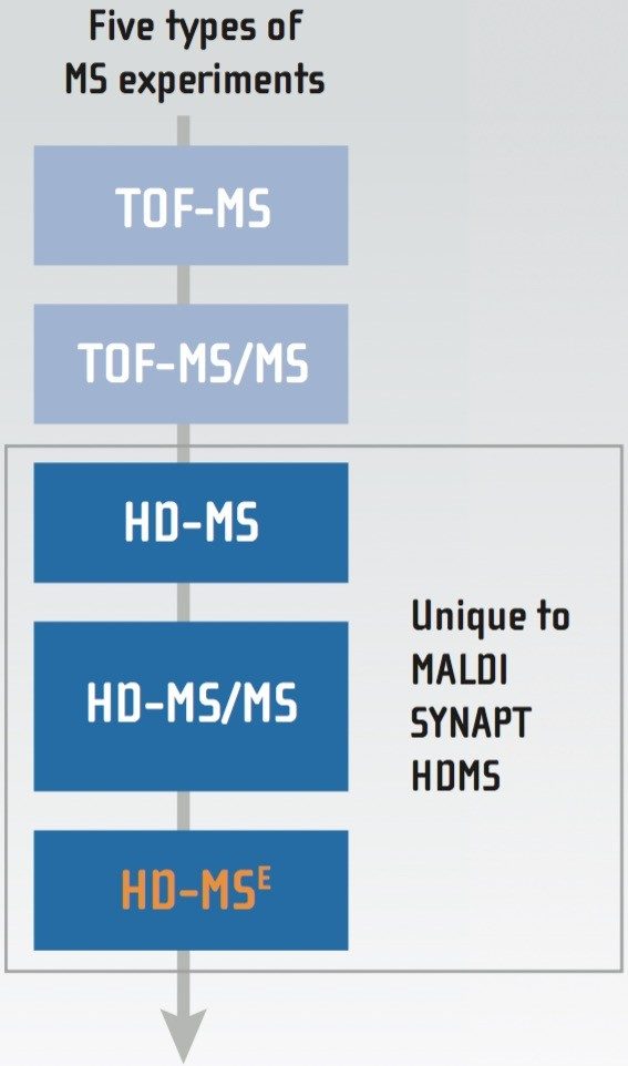 Figure 1. Experiments supported using Waters HDI Imaging Software.
Workflow of the HDI Software
The initial step is to use the pattern definition tool to assign reference points to the photographic image, in order to select the area of interest for HD imaging.
The processing experiment file is created directly from the HDI Software and loaded into a MassLynx™ Software sample list. Processing of the data using the algorithm Apex 3D to create a peak list with m/z and drift time information occurs automatically after acquisition.
The resulting raw data are processed in the Analysis Section of the software, where all types of experiments described in Figure 1 are supported. Analysis of the acquired imaging data sets fully incorporates the ion mobility information, which is integrated into the data processing and visualization, as shown in Figure 2. This allows the distribution of molecules such as drugs, lipids, or peptides to be determined without the interference of background ions or isobaric species.
Smooth interactions between the available visualizations – including the peak list table, mass spectrum, drift time versus m/z plot – and the ion images allow scientists to analyze their data in a powerful, user-friendly fashion.
Following fully automated HDI data acquisition and processing, the results can be exported as raw data for statistical treatment using MarkerLynx™ XS Application Manager, or directly into other MassLynx applications for further processing, such as the elemental composition tool, or MassFragment™.
The user-defined grid gallery allows the comparison of a series of ion images of interest by using the Red/Green/Blue (RGB) overlay capability.
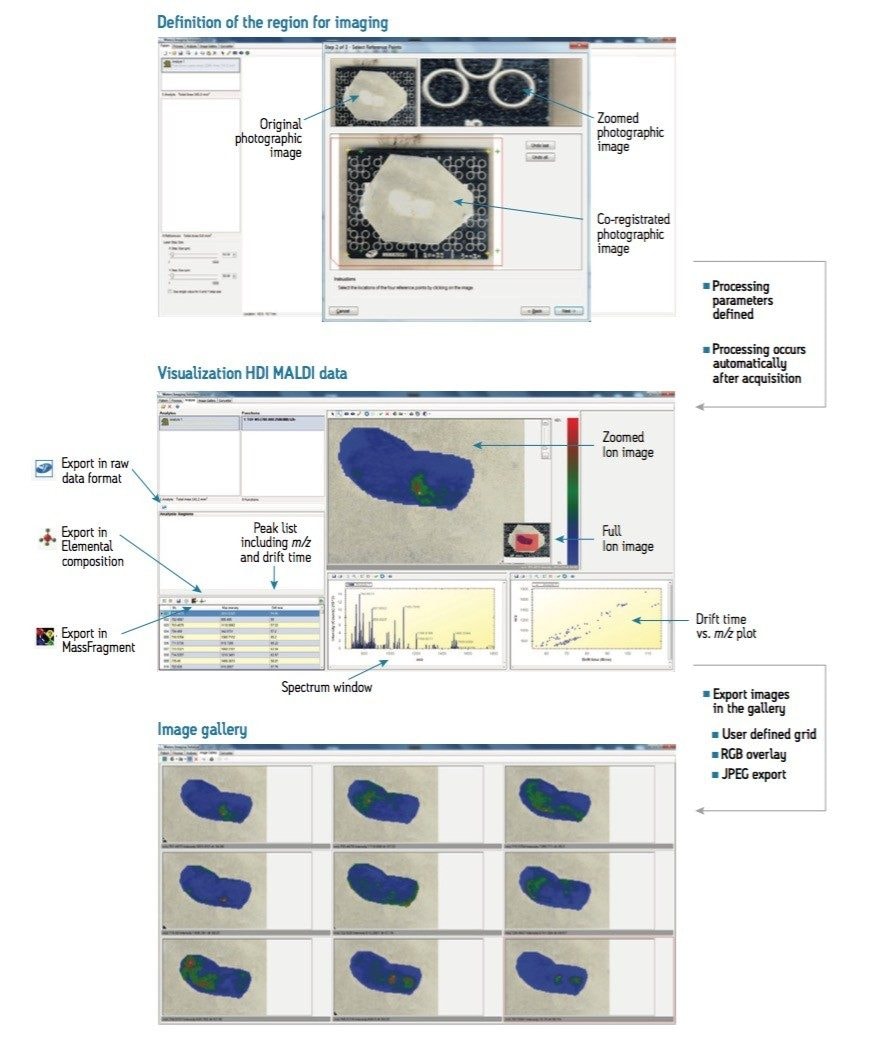 Figure 2. Acquired imaging data sets fully incorporate the ion mobility information that is integrated into the data processing and visualization tool.
Conclusion
- Waters’ High Definition Imaging Software is a new, fully integrated software suite for MALDI imaging experiments
- Integration of HDI data acquisition processing and visualization is performed in a single interface
- For the first time, MALDI imaging ion mobility information is fully incorporated and used within the imaging software
- MALDI HDMSE data can be acquired and easily visualized
- Flexible export options are available for calculating elemental composition, statistical analysis using MarkerLynx XS Application Manager, or analysis with MassFragment
720003988, May 2011