Evaluation of Modern and Legacy HPLC Systems for the Size-Exclusion Analysis of Aggregation and Fragmentation in a Monoclonal Antibody Drug
Abstract
Size-exclusion chromatography is a widely used characterization technique for the analysis of aggregation and fragmentation of monoclonal antibody drugs. Traditional high-performance liquid chromatography systems have a large amount of extra-column dispersion which makes size-exclusion data analysis difficult due to band broadening and increased peak tailing. Additionally, biologic analytes may experience non-specific adsorption throughout the chromatographic flow path, further increasing the effects of peak tailing. The Alliance™ iS Bio HPLC System addresses both concerns by offering lower system dispersion than legacy HPLC systems alongside a bio-inert flow path employing MaxPeak™ High-Performance Surfaces Technology.
In this work an Alliance iS Bio HPLC System, an Alliance e2695, and a competitive Bio-inert HPLC system are compared using a size-exclusion method for analysis of monoclonal antibody aggregation and fragmentation. Dispersion on each system is determined and the Alliance iS Bio HPLC is shown to have the lowest 4σ dispersion. This translates to lower peak tailing, tighter peak width, and a more accurate measurement of low-molecular weight species of infliximab due to lower monomer peak tailing and better monomer resolution.
Benefits
- Biocompatible system for use in salty bio-applications
- MaxPeak HPS for reduced off-target analyte interaction results in less peak tailing
- Low system dispersion results in better fragmentation determination
Introduction
Characterization of monoclonal antibodies (mAbs) requires a multi-pronged approach targeting specific characteristics of the large molecule drug product. From a top-down perspective, looking at the intact protein via size-exclusion chromatography (SEC) can be useful for monitoring aggregation or fragmentation of the main drug product. Aggregate formation can occur during the fermentation, purification, and storage steps of the manufacturing process and may lead to immunogenic reactions or loss of drug efficacy.1 Fragmentation of the drug product can also occur during the formulation process either spontaneously or enzymatically, and is an indicator of the stability of the product.2 Both aggregation and fragmentation are considered critical quality attributes (CQAs) and must be monitored throughout the drug lifecycle.
SEC is the gold standard for monitoring aggregation and fragmentation of mAbs. This technique uses a buffered mobile phase system at a consistent pH to limit off-target interactions of the analyte with the chromatographic flow path, allowing for a separation based on the hydrodynamic radii of the analytes. However, this assumes that the buffering system in the mobile phase is ideal for all analytes in solution. Practically, this is not always true and interactions between analytes and the chromatographic flow path do occur. This problem is particularly tricky when working with large molecule biologics. mAbs are large proteins (roughly 150 kDa) with various positively and negatively charged patches found on the surface of their 3-dimensional structure. These charged patches tend to interact with charged surfaces in the flow path leading to reduced chromatographic quality in the form of peak tailing.
In recent years biocompatible chromatography systems have been introduced which use non-ferrous materials such as titanium, PEEK, and MP35N throughout the flow path to prevent corrosion due to the salty buffers often used in biological analyses. Waters™ has recently introduced the Alliance iS Bio HPLC System, which incorporates MaxPeak High-Performance Surfaces (HPS) Technology alongside a bioinert flow path. The HPS technology acts as a barrier to off-target interactions between analytes and the flow path, improving chromatographic quality for molecules that tend to interact. In this study the Alliance iS Bio HPLC System is compared to two legacy HPLC systems for use in the SEC analysis of infliximab for monitoring aggregation and fragmentation levels.
Experimental
Sample Description
BEH™ 200 SEC Protein Standard Mix (p/n: 186006518) was reconstituted in 500 µL of mobile phase, vortexed, and passed through a 0.2 µm filter.
Remicade™ (infliximab) was diluted to 10 mg/mL with water. 500 µL aliquots were stored at -80 °C until needed. The drug product used in this study was analyzed past expiry.
LC Conditions
|
LC system: |
Alliance iS Bio HPLC System Alliance e2695 HPLC System Competitive Bio-Inert HPLC System (System Y) |
|
Detection: |
UV detection: 280 nm @ 10 points/second |
|
Vials: |
Maximum Recovery (p/n: 186005662CV) for IEX Cation Test Standard Total Recovery (p/n: 186005663CV) for Infliximab |
|
Column: |
XBridge™ Premier Protein SEC, 4.6 x 300 mm, 250 Å, 2.5 µm (p/n: 186009960) |
|
Column temperature: |
Ambient |
|
Sample temperature: |
6 °C |
|
Injection volume: |
5 µL for BEH200 SEC Protein Standard Mix 10 µL for Infliximab |
|
Flow rate: |
0.3 mL/min |
|
Mobile phase: |
60.3 mM dibasic potassium phosphate, 140.3 mM monobasic potassium phosphate, 249.5 mM potassium chloride @ pH 6.2. Pass mobile phase through a 0.2 µm filter. |
|
Gradient: |
Isocratic |
Data Management
|
Chromatography software: |
Alliance iS Bio : Empower™ 3.8.0.1 Alliance e2695 : Empower 3.7 System Y: Empower 3.7 |
Results and Discussion
System dispersion can have a large impact on the quality of SEC results as larger dispersion values lead to wider peaks and increased chromatographic tailing. The implications of system dispersion, SEC column choice, and separation conditions have been discussed previously.3–4 Pre-column dispersion is as problematic as post-column dispersion in SEC due to the lack of sample re-focusing at the head of the column. The Alliance iS Bio HPLC System delivers the lowest system dispersion of the HPLC systems tested (Table 1). The implications of system dispersion can be observed using the BEH200 SEC Protein Standard Mix. (Figure 1).
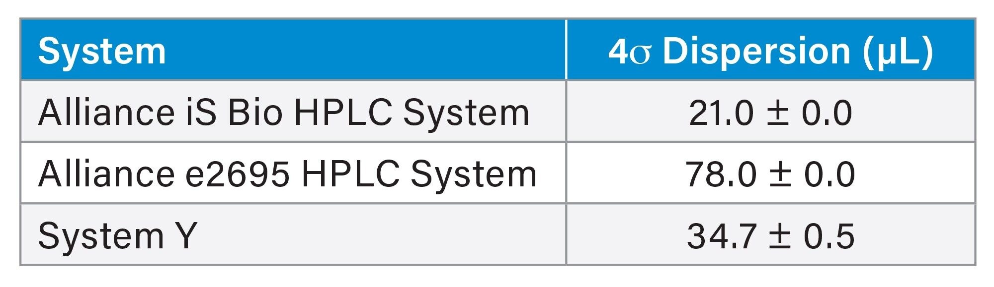 Table 1. 4σ dispersion measurements given in µL for the 3 HPLC systems tested in this study.
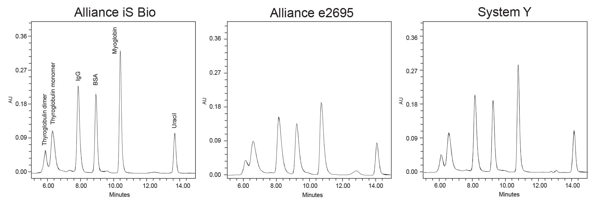 Figure 1. Overlayed duplicate injections of BEH200 SEC Protein Standard Mix on each of the three systems. Compounds are labeled in the left-most frame.
The Alliance iS Bio and System Y demonstrated lower system dispersion and peak tailing measurements compared to the Alliance e2695. (Table 2) Analyte peaks on the Alliance e2695 are broader and demonstrate lower peak heights in part due to increased system dispersion. As the analyte bands pass through the column and post-column/pre-detector tubing turbulent flow along with natural Brownian motion cause the molecules to diffuse and therefore dilute. Systematic differences in fittings and tubing diameter also play a role in system dispersion. The decrease in peak tailing observed on the biocompatible Alliance iS Bio System and bio-inert System Y may be attributed to the materials used throughout the flow path of the system. As the biomolecules pass through the flow path there is a chance that they interact with exposed charged metal surfaces throughout resulting in increased tailing of the analyte peak. The Alliance e2695 materials are more susceptible to these interactions and the resulting increase in peak tailing may be the result.
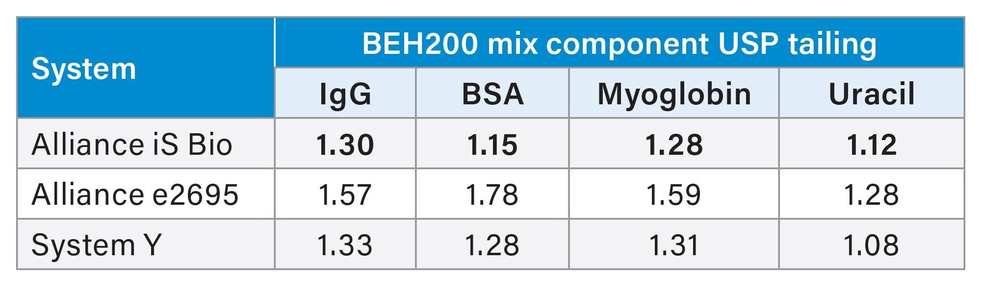 Table 2. USP peak tailing measurements for the final four compounds in the BEH200 SEC Protein Standard Mix on each system. Results are the mean of two injections.
The effects of increasing system dispersion and peak tailing can be observed with a more difficult separation such as looking at high-molecular weight species (HMWS) and low-molecular weight species (LMWS) in a mAb drug. (Figure 2) The HMWS are dimers and higher-level self-associated or aggregate formations of the monomer, while LMWS are fragmented parts of the monomer. System Y and the Alliance e2695 demonstrated higher peak tailing than the Alliance iS Bio System for this separation.
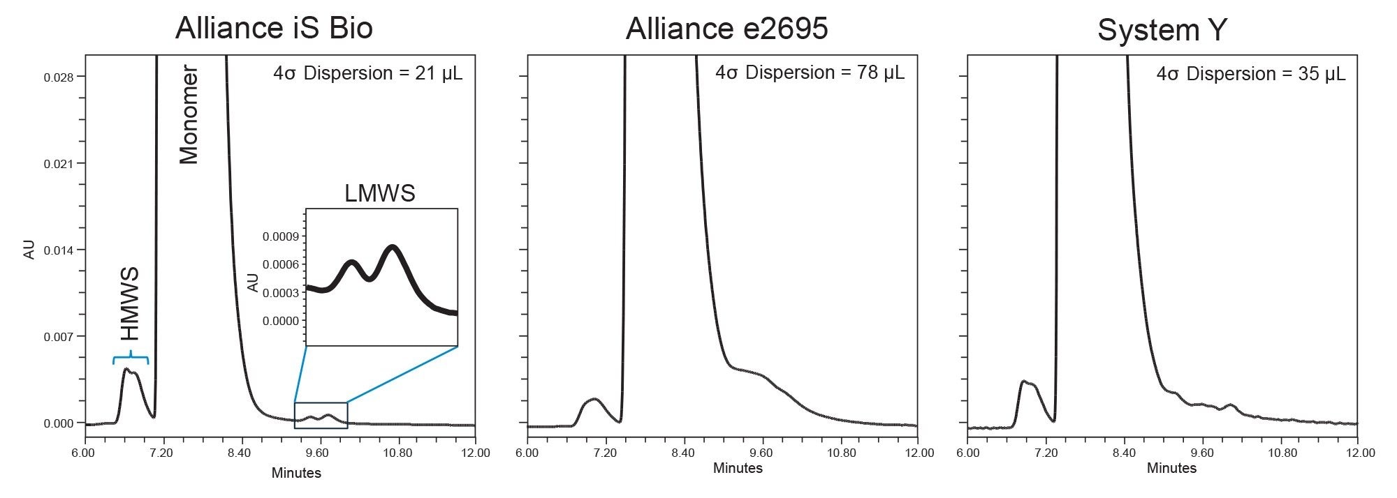 Figure 2. A single injection of the monoclonal antibody infliximab on each system. The y-axis of each chromatogram is scaled to the left-most frame. HMWS=high-molecular weight species. LMWS=low-molecular weight species.
Peak tailing of the monomer causes the area counts of the poorly resolved and minute LMWS peaks to be artificially increased in the case of System Y while they are completely buried by the monomer peak tail on the Alliance e2695. When calculating LMWS in each of these cases there is ambiguity as to where the correct placement of the peak start should be. In this instance, the tailing mound on the Alliance e2695 was included in the monomer peak area, however if the analyst were to integrate the tail as the fragment, the amount of LMWS would be greatly overestimated. The Alliance iS Bio System was the only system tested which nearly returns to baseline prior to elution of the LMWS peaks, resulting in a truer measure of the fragmentation peaks in solution. (Table 3) In addition to the lower dispersion on the Alliance iS Bio System compared to System Y, the improved peak tailing and LMWS detection may also be attributed to the HPS modification used throughout the system. Both systems are designed for use in bio-applications, but with the HPS potentially providing additional adsorption protection the monomer may demonstrate better peak shape. On the leading edge of the monomer peak, the HMWS on each system is relatively well resolved and relative area (% Area) measurements are comparable between systems. (Table III) The broader peaks of the Alliance e2695 along with the increase in tailing on this system do however impact separation of the HMWS from the monomer in terms of peak/valley (P/V) measurements. Ending P/V of the HMWS peak was 6.6, 8.0, and 8.1 for the Alliance e2695, System Y, and the Alliance iS Bio System respectively.
 Table 3. Relative area measurements from a single injection of Infliximab on all systems.
Conclusion
The Alliance iS Bio HPLC System offers low system dispersion alongside a biocompatible flow path with MaxPeak HPS technology and is well suited for use in bioanalytical methods. When compared with an Alliance e2695 and a competitive bio-inert system (System Y), the lower system dispersion translates to reduced peak width as a result of decreased extra-column band broadening. When each system was used for monitoring aggregation and fragmentation of a mAb, the increased peak tailing observed on the Alliance e2695 and System Y leads to difficulties when analyzing fragmentation peaks. The Alliance iS Bio System is readily able to distinguish the low-level (0.20% relative area) fragment peaks from the tail of the abundant monomer peak, while the Alliance e2695 completely loses these peaks in the monomer tail and System Y exaggerates their abundance. The larger system dispersion of the Alliance e2695 also contributed to decreased separation of the aggregate species from the monomer peak. With fragmentation and aggregation level monitoring being a CQA for the analysis of mAb biologics, a clear picture of the levels of these impurities is required, and the Alliance iS Bio System outperforms comparable HPLC systems in this regard.
References
- Vázquez-Rey, M.; Lang, D. A. Aggregates in Monoclonal Antibody Manufacturing Processes. Biotechnol. Bioeng. 108 (7), 1494–1508. https://doi.org/10.1002/bit.23155. 2011.
- Vlasak, J.; Ionescu, R. Fragmentation of Monoclonal Antibodies. mAbs 3 (3), 253–263. https://doi.org/10.4161/mabs.3.3.15608. 2011.
- Koza, S. M.; Reed, C.; Chen, W. Evaluating the Impact of LC System Dispersion on the Siz-Exclusion Chromatography Analysis of Proteins. 2019.
- Koza, S. M.; Reed, C. E.; Chen, W. Impact of LC System Dispersion on the Size-Exclusion Chromatography Analysis of Monoclonal IgG Antibody Aggregates and Fragments: Selecting the Optimal Column for Your Method. Waters Application Note 720006336. 2019.
720008614, December 2024