• Note d'application

Using Hydrophilic Interaction Chromatography for Heightened Product Characterization to Overcome Challenges with Hydrophobic Monoclonal Antibodies and Antibody Drug Conjugates

Using Hydrophilic Interaction Chromatography for Heightened Product Characterization to Overcome Challenges with Hydrophobic Monoclonal Antibodies and Antibody Drug Conjugates

  • Jacquelynn Smith
  • Matthew A. Lauber
  • Stephan M. Koza
  • Erin E. Chambers
  • Jason C. Rouse
  • Olga V. Friese
  • Biotherapeutics Pharm. Sci., Pfizer Inc
  • Waters Corporation

Abstract

Hydrophilic interaction chromatography (HILIC) is a compelling alternative to reversed-phase liquid chromatography (RPLC) for addressing challenges that have long been encountered with conventional mAb separation modes. The hydrophobic subunits of monoclonal antibodies and antibody drug conjugates can, for instance, be challenging to completely recover by RPLC because of their tendency to undergo disadvantageously strong analyte-to ligand adsorption. For the molecules tested here, HILIC yielded considerably higher recovery than RPLC without signs of on-column hydrolysis. Both mAb and ADC subunits were effectively separated. And with inherent MS compatibility, the HILIC method was easily hyphenated with MS allowing for identifications to be made via accurate on-line mass analyses. In sum, HILIC has been found to be complementary to RPLC and a promising new tool for biotherapeutic protein characterization.

Benefits

  • Complementary, orthogonal separations to RPLC
  • Improved recovery of hydrophobic protein components
  • Reduced temperature dependence for decreasd on-column degradation

Introduction

While hydrophilic interaction chromatography (HILIC) has been widely used for separating small polar compounds, its application to large biomolecules, other than released glycans, has been surprisingly limited. In large part, this can be attributed to a paucity of suitable HILIC column technology and robust separation methods. This is despite the potential for HILIC to be highly valuable for the characterization of whole proteins with and without glycosylation. IgG based monoclonal antibodies (mAbs) have two identical heavy chains and two identical light chains that are held together by both covalent disulfide bonds and non-covalent interactions. More importantly, mAbs exhibit two functionally significant subunits: two equivalent antigen binding fragments (Fab domains), and one crystallizable fragment (Fc domain), which is glycosylated with N-linked complex-type biantennary structures. Structural characterization of these subunits typically involves proteolysis by the Immunoglobulin Degrading Enzyme of S. pyogenes (IdeS) (Figure 1). IdeS has high specificity for a conserved Gly-Gly sequence motif in the lower hinge region of mAbs, and when used with disulfide bond reduction, IdeS produces three different 25 kDa mAb fragments: light chain (LC), along with two heavy chain Fd' and single chain Fc (scFc) domains.1 Normally, characterization of these three antibody constituents is achieved by reversed-phase liquid chromatography (RPLC) coupled to ultrahigh-resolution mass spectrometry (MS). Many times, it can be difficult to achieve complete chromatographic recovery of all three subunits/domains due to their hydrophobic nature. And while ultra-high temperatures can improve recoveries, such RPLC method conditions can become problematic because of analyte degradation and the potential to introduce method artifacts.

Figure 1. IdeS digestion and reduction scheme for preparing IgG LC, Fd', and scFc subunits.

Hydrophilic interaction chromatography appears to be a promising alternative to RPLC for mAb subunit analysis. Here, a recently introduced stationary phase constructed of amide-bonded, wide-pore organosilica particles has been used to perform separations that are complementary to RPLC and produce improved chromatographic recovery of hydrophobic mAb and antibody drug conjugate (ADC) subunits.

Experimental

Sample description

IdeS digestion and reduction of mAbs:

The therapeutic mAb and ADC used in this study were manufactured by Pfizer, Inc. Treatment with IdeS (Genovis AB, Lund, Sweden), an enzyme that results in lower hinge region proteolysis, followed by disulfide bond reduction, was used to generate mAb light chain and the heavy chain scFc and Fd' subunits.

For example, the therapeutic mAb was diluted to 2 mg/mL (50 μL total) in digestion buffer (pH 6.6), mixed with 2 μL of IdeS enzyme (approximately 118 U), and incubated at 37 °C for 1 hour. To denature and reduce the resulting subunits, 100 μL of 8 M guanidine, 25 μL of IdeS digestion buffer, and 25 μL 1 M DTT were added to the sample, followed by incubation at 37 °C for 90 minutes.

Intact mAb Mass Check Standard, a mouse monoclonal IgG1, was obtained from Waters (p/n 186006552).

Method conditions (unless otherwise noted):

Column conditioning

ACQUITY UPLC Glycoprotein BEH Amide, 300Å, 1.7 μm columns were conditioned via repeating high mass load injections of the mAb until consistent peak shape was observed.

LC condition (Figures 2 and 4):

LC system:

ACQUITY UPLC H-Class System

Sample temp.:

8 °C

Flow rate:

0.2 mL/min

Injection volume:

10 μL (RPLC), 2.5 μL (HILIC)

Sample load:

5 μg (RPLC), 1.3 μg (HILIC)

Columns:

ACQUITY UPLC Glycoprotein BEH Amide, 300A, 1.7 μm, 2.1 x 150 mm, (p/n 176003702, with Glycoprotein Performance Test Standard)

ACQUITY UPLC Protein BEH C4, 300A, 1.7 μm, 2.1 x 100 mm (p/n 186004496)

Temperature:

As noted

UV detection:

A214

HILIC chromatographic conditions

Mobile phase A:

100% H2O (v/v)

Mobile phase B:

100% ACN (v/v)

Mobile phase C:

2% TFA (v/v)

Gradient

Time

%A

%B

%C

Curve

0.00

20.0

77.5

2.5

6

1.00

20.0

77.5

2.5

6

21.0

34.5

63.0

2.5

6

22.0

97.5

0.00

2.5

6

24.0

97.5

0.00

2.5

6

25.0

20.0

77.5

2.5

6

35.0

20.0

77.5

2.5

6

RPLC chromatographic conditions

Mobile phase A:

100% H2O (v/v)

Mobile phase B:

100% ACN (v/v)

Mobile phase C:

2% TFA (v/v)

Mobile phase D

80:20 IPA:ACN (v/v)

Gradient

Time

%A

%B

%C

%D

Curve

0.0

87.4

5.7

5.0

1.9

6

3.0

87.4

5.7

5.0

1.9

6

5.0

71.2

11.3

5.0

12.5

6

45.0

52.2

17.8

5.0

25.0

6

47.0

4.7

34.1

5.0

56.2

6

53.0

4.7

34.1

5.0

56.2

6

53.1

87.4

5.7

5.0

1.9

6

54.0

4.7

34.1

5.0

56.2

6

55.0

87.4

5.7

5.0

1.9

6

62.0

87.4

5.7

5.0

1.9

6

LC condition (Figure 3)

LC system:

ACQUITY UPLC H-Class Bio System

Sample temp.:

10 °C

Flow rate:

0.2 mL/min

Injection volume:

0.5 μL (2 mg/mL Intact mAb Mass Check Standard)

Columns:

ACQUITY UPLC Glycoprotein BEH Amide, 300A, 1.7 μm, 2.1 x 150 mm, (p/n 176003702, with Glycoprotein Performance Test Standard)

ACQUITY UPLC Protein BEH C4, 300A, 1.7 μm, 2.1 x 150 mm (p/n 186004497)

Mobile phase A:

0.1% (v/v) TFA, H2O

Mobile phase B:

0.1% (v/v) TFA, ACN

Temperature:

As noted

UV detection

A214

HILIC chromatographic conditions

Time

%A

%B

Curve

0.0

20.0

80.0

6

1.0

32.0

68.0

6

21.0

39.0

61.0

6

22.0

80.0

20.0

6

24.0

80.0

20.0

6

25.0

20.0

80.0

6

35.0

20.0

80.0

6

RPLC chromatographic conditions

Time

%A

%B

Curve

0.0

95.0

5.0

6

1.0

62.0

38.0

6

21.0

55.0

45.0

6

22.0

20.0

80.0

6

24.0

20.0

80.0

6

25.0

95.0

5.0

6

35.0

95.0

5.0

6

Results and Discussion

HILIC for large biomolecules

The use of HILIC for the characterization of protein biopharmaceuticals has been made possible by a new sub-2 μm stationary phase constructed of amide-bonded, wide-pore organosilica particles. This phase facilitates separations of unconventionally large analytes because it has a sufficiently large average pore diameter that allows proteins to have enhanced access to the bonded phase.2 Previous work with this stationary phase focused on its ability to resolve the glycoforms of proteins, protein subunits and peptides.3-5 With this work, mAb and ADC subunit separations achieved by HILIC versus RPLC were compared from a different perspective, allowing additional benefits of HILIC to be revealed.

Hydrophobic mAb subunits

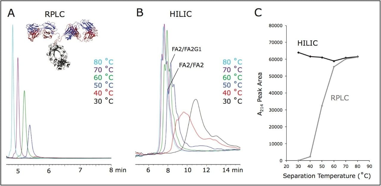
Many times, the hydrophobic nature of a protein requires that RPLC be performed with high column temperatures in order to ensure quantitative chromatographic recovery.6 Unfortunately, these conditions can sometimes cause on-column hydrolysis, particularly when it is necessary to use shallow, prolonged gradients to resolve subunits with similar polarities. In addition, it may actually be impractical to achieve complete chromatographic recovery of all species originating from an inordinately hydrophobic mAb.

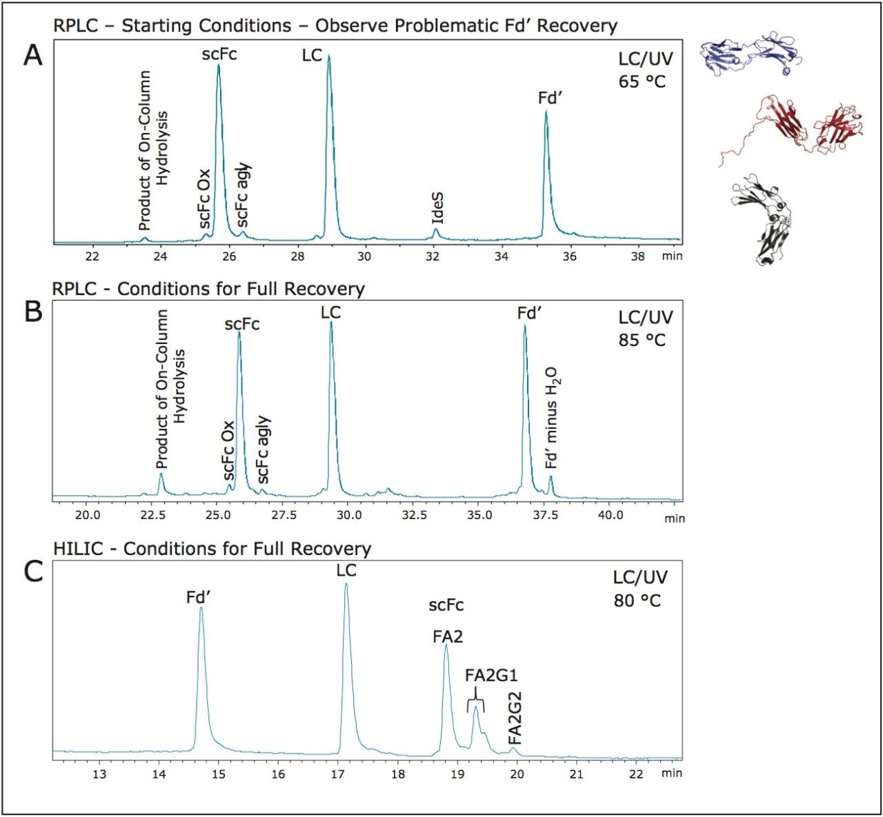
Figure 2 demonstrates an example in which RPLC led to a compromised analysis of subunits from a therapeutic mAb. For this RPLC separation, method development started with a recommended temperature of 65 °C and a TFA level of 0.1% (Figure 2A). However, these conditions resulted in noticeably poor recovery of the Fd' subunit. Additional method optimization revealed that it was necessary to use 0.1% TFA combined with an 85 °C column temperature to ensure quantitative recovery of the Fd' subunit (Figure 2B). While the RPLC chromatogram obtained at 85 °C shows sharp, abundant peaks for each of the three mAb subunits, it also shows evidence of on-column degradation, as indicated by the peak with a retention time of 22.9 minutes. Analysis of this peak by online MS and accurate mass measurement has shown it to be a result of hydrolytic cleavage at a labile amide bond between an aspartic acid and proline residue. Along with this prominent artifact peak, it can be seen that the baseline of the obtained RPLC chromatogram is populated with additional spurious peaks.

A method with a susceptibility to generating artifacts can prove to be challenging to implement due to ambiguous data interpretation. For this reason, HILIC was investigated as an alternative means of chromatographically profiling these mAb subunits. It was found that both optimal resolution and recovery of the mAb subunits could be obtained with this mode of separation using 0.05% TFA and a column temperature of no more than 80 °C. As is displayed in Figure 2C, the HILIC chromatogram that was obtained for the mAb subunits presented more than three peaks. The first two peaks correspond to the Fd' and LC subunits, while the last grouping of peaks can be attributed to the glycoforms of the scFc subunit. This is an elution order that, as predicted, is the reverse of that generated by RPLC.

It is clear then that HILIC and RPLC separations are orthogonal. This is reasonable as their retention mechanisms are entirely different. Protein retention via RPLC involves hydrophobic adsorption and an ‘on/off’ separation with relatively limited partitioning. In contrast, HILIC, of even proteins, is believed to involve partitioning of an analyte into an immobilized water layer, where it can undergo hydrogen bonding, dipole-dipole, and ionic interactions with the stationary phase.7 Not only does this mean that orthogonal separations can be obtained, it suggests that the interaction between a protein and a stationary phase may be more reversible in HILIC than RPLC. Indeed, Figure 2C indicates that HILIC can show more favorable recovery than RPLC for a hydrophobic Fd' subunit.


Figure 2. Comparison of RPLC and HILIC for mAb subunit analysis demonstrating separations that yielded both partial (A) and full recovery (B, C) of the Fd' subunit. Glycosylation is labeled according to Oxford notation. F denotes fucose, A2 represents that the structures are biantennary, and G stands for galactose (FA2 = G0F, FA2G1 = G1F, and FA2G2 = G2F). 'Ox' and 'agly' denote oxidation and aglycosylated, respectively.

Interestingly, the HILIC separation of these mAb subunits also seems to be conspicuously free of the on-column degradation product that was otherwise observed during RPLC-based profiling. While the nature of this phenomenon has not been explored, it is perhaps due in part to the combined effect of a lower temperature, lower TFA concentration, and a mobile phase composition that is comparatively lower in water. Additionally, the nature of protein unfolding on the reversed-phase based hydrophobic surfaces may also be a contributing factor.8

Recovery as a function of temperature – HILIC versus RPLC

Given the significance of the above observations, we aimed to more precisely define the temperature dependence of HILIC versus RPLC. An intact monoclonal IgG1 antibody, specifically the Waters Intact mAb Mass Check Standard, was separated by either RPLC or HILIC using identical mobile phases along with column temperatures ranging from 30 up to 80 °C. Chromatograms corresponding to these analyses are displayed in Figures 3A and 3B. As predicted, mAb recovery from the RPLC column improved with increasing separation temperatures, an observation consistent with reports on the problematically strong adsorption of mAbs and their fragments to reversed-phase sorbents.6 HILIC, in contrast, produced high recoveries of the mAb regardless of the separation temperature. These findings are most apparent in plots that show peak area as a function of temperature (Figure 3C). Notice that HILIC generated a peak area at 50 °C that was equivalent to that obtained at 80 °C. Meanwhile, when RPLC was employed at 50 °C, only 53% of the peak area was calculated to be recovered compared to results obtained at 80 °C. These data thereby indicated that a 50 °C separation is optimal for HILIC of this intact mAb (Figure 3B, blue trace). In fact, at such a temperature, the HILIC separation exhibited sufficient selectivity and resolution to partially resolve individual glycoforms4 (FA2/FA2 versus FA2/FA2G1; Oxford notation9).

Figure 3. Temperature dependence of HILIC versus RPLC. Chromatograms of intact mAb mass check standard (mouse monoclonal IgG1) as obtained at various temperatures using RPLC (A) or HILIC (B). UV peak area as a function of temperature (C). Glycosylation is labeled according to Oxford notation. F denotes fucose, A2 represents that the structures are biantennary, and G stands for galactose (FA2 = G0F, FA2G1 = G1F, and FA2G2 = G2F).

These observations confirm that HILIC can indeed show a significantly lower dependence on temperature than RPLC. Remarkably high recovery can be achieved with HILIC at unconventionally low column temperatures, which could be advantageous in developing LC profiling techniques for particularly challenging molecules that are either extraordinarily hydrophobic or susceptible to temperature-accelerated degradation.

Higher recovery ADC subunit analysis

That HILIC facilitates the chromatographic recovery of hydrophobic species could lend itself to the analysis of ADCs. Although there are only two ADC products currently available on the market, Kadcyla and Adcetris, there are many more in pre-clinical and clinical stages of development.10-11

IdeS-based subunit analyses of ADCs are frequently used to fingerprint the extent of conjugation and to quickly assess domain specific information about the linkages.

However, the hydrophobicity of the so-called linker/drug ‘payloads,’ including auristatin and maytansinoid conjugates, can make it challenging to obtain proper chromatographic recovery of all subunits/domains. Also, many ADCs have very hydrophobic heavy chains and Fd' sequences, in particular. The conjugation of these hydrophobic payloads onto already hydrophobic moieties within mAbs can therefore confound an RPLC analysis. Nevertheless, RPLC is a standard platform separation for such an analysis, given its potential for high resolution and compatibility with MS detection.

Interestingly, separations of a prototypical ADC subunit sample suggest that HILIC is a compelling alternative (Figure 4). As expected, the HILIC separation of ADC subunits exhibited orthogonality to RPLC. Not surprisingly, the addition of the hydrophobic payload to a subunit leads to a decrease in retention, which is contrary to reversed-phase behavior. While resolution does not appear to be as high as with RPLC, HILIC does nevertheless show an ability to resolve individual positional isomers (i.e., Fd' 1a versus Fd' 1b). Most importantly and as shown in the displayed profiles, quantitative recovery of the Fd', LC, and scFc subunits was obtained with HILIC for both the precursor mAb and its corresponding ADC. Specifically, the sum of the percent peak areas for the various Fd' ADC species (35.0%) is in good agreement with the percent peak area of the Fd' subunit from the precursor mAb (36.7%). Accordingly, the addition of the payload appeared to have minimal, if any impact, on the recovery of the subunits from the HILIC chromatography.

Figure 4. Hydrophilic Interaction Chromatography of a mAb versus its ADC. Numbers represent how many linker/payloads are present in the conjugate, where a and b denote positional isomers. *Denotes linker/payload hydrolysis products. Denotes trace level oxidation. Glycosylation is labeled according to Oxford notation. F denotes fucose, A2 represents that the structures are biantennary, and G stands for galactose (FA2 = G0F, FA2G1 = G1F, and FA2G2 = G2F).

Conclusion

Hydrophilic interaction chromatography (HILIC) is a compelling alternative to reversed-phase liquid chromatography (RPLC) for addressing challenges that have long been encountered with conventional mAb separation modes. The hydrophobic subunits of monoclonal antibodies and antibody drug conjugates can, for instance, be challenging to completely recover by RPLC because of their tendency to undergo disadvantageously strong analyte-to-ligand adsorption. Because HILIC is achieved by means of an entirely different retention mechanism, hydrophobic protein subunits do not strongly adsorb to the HILIC stationary phase and can, as a result, be eluted with less extreme conditions.

For the molecules tested here, HILIC yielded considerably higher recovery than RPLC without signs of on-column hydrolysis. Both mAb and ADC subunits were effectively separated. And with inherent MS compatibility, the HILIC method was easily hyphenated with MS allowing for identifications to be made via accurate on-line mass analyses. In sum, HILIC has been found to be complementary to RPLC and a promising new tool for biotherapeutic protein characterization. While HILIC of mAb subunits can be used to assess N-glycosylation profiles, this study shows that it can also be taken advantage of to more comprehensively recover the subunits of mAbs and ADCs.

References

  1. An, Y.; Zhang, Y.; Mueller, H.M.; Shameem, M.; Chen, X. A new tool for monoclonal antibody analysis: application of IdeS proteolysis in IgG domain specific characterization. MAbs 2014, 6 (4), 879–93.
  2. Renkin, E.M. J. Gen. Physio. 1954, (38), 225.
  3. Lauber, M.A.; Koza, S.M. Mapping IgG Subunit Glycoforms using HILIC and a Wide-Pore Amide Stationary Phase. Waters Application Note 720005385EN, 2015.
  4. Lauber, M.A.; Koza, S.M. Developing High Resolution HILIC Separations of Intact Glycosylated Proteins using a Wide-Pore Amide-Bonded Stationary Phase. Waters Application Note 720005380EN, 2015.
  5. Lauber, M.A.; Koza, S.M. HILIC Glycopeptide Mapping with a Wide-Pore Amide Stationary Phase. Waters Appication Note 720005409EN, 2015.
  6. Fekete, S.; Beck, A.; Wagner, E.; Vuignier, K.; Guillarme, D. Adsorption and recovery issues of recombinant monoclonal antibodies in reversed-phase liquid chromatography. J. Sep. Sci. 2015, 38 (1), 1–8.
  7. Alpert, A.J. Hydrophilic-interaction chromatography for the separation of peptides, nucleic acids and other polar compounds. J. Chromatogr. 1990, 499, 177–96.
  8. McNay, J.L.; Fernandez, E.J. How does a protein unfold on a reversed-phase liquid chromatography surface? J. Chrom. A 1999, 849 (1), 135–148.
  9. Oxford Notation: FA2 = GOF, FA2G1 = G1F. F denotes fucose, A2 represents a biantennary structure, and G stands for galactose.
  10. Casi, G.; Neri, D. Antibody-Drug Conjugates and Small Molecule-Drug Conjugates: Opportunities and Challenges for the Development of Selective Anticancer Cytotoxic Agents. J. Med. Chem. 2015, 58 (22), 8751–61.
  11. Shen, W.-C. Antibody-Drug Conjugates: A Historical Review. In Antibody-Drug Conjugates: The 21st Century Magic Bullets for Cancer, Shen, W.-C.; Wang, J.; Zaro, J.L., Eds. 2015.

720005684, April 2016

Haut de la page Haut de la page