As an initial step towards transferring peptide mapping methods from HPLC to UPLC, we previously presented an approach using the ACQUITY UPLC H-Class Bio System for legacy HPLC-based peptide mapping.1 Our method transfer discussion continues here, focusing on improving the consistency of peptide mapping separations during routine analyses.
Peptide mapping methods generally include an acidic modifier to improve peak shape. However, accurate and reproducible management of the modifier content within mobile phase solvents can be variable, consequently affecting the peptide map quality and reproducibility.
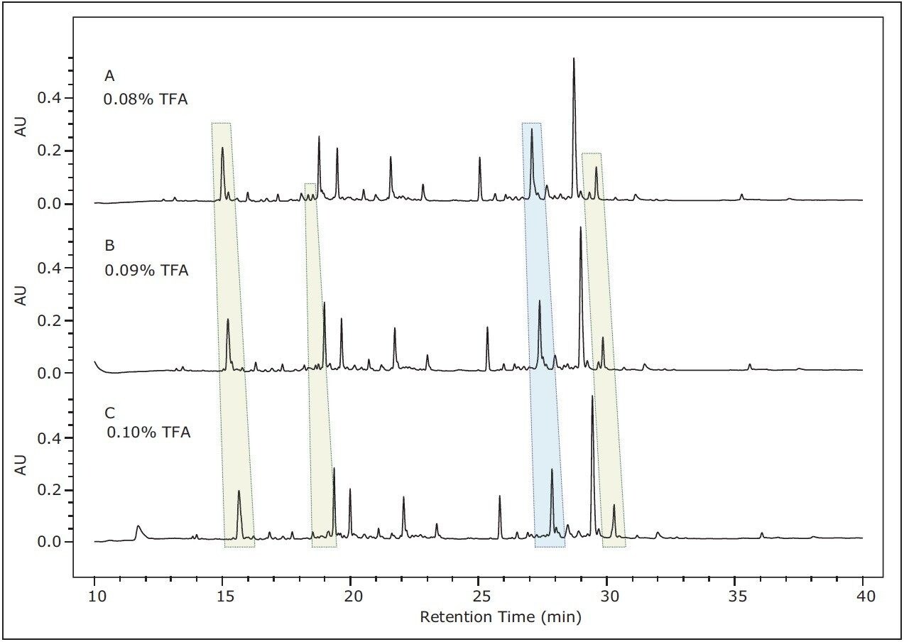
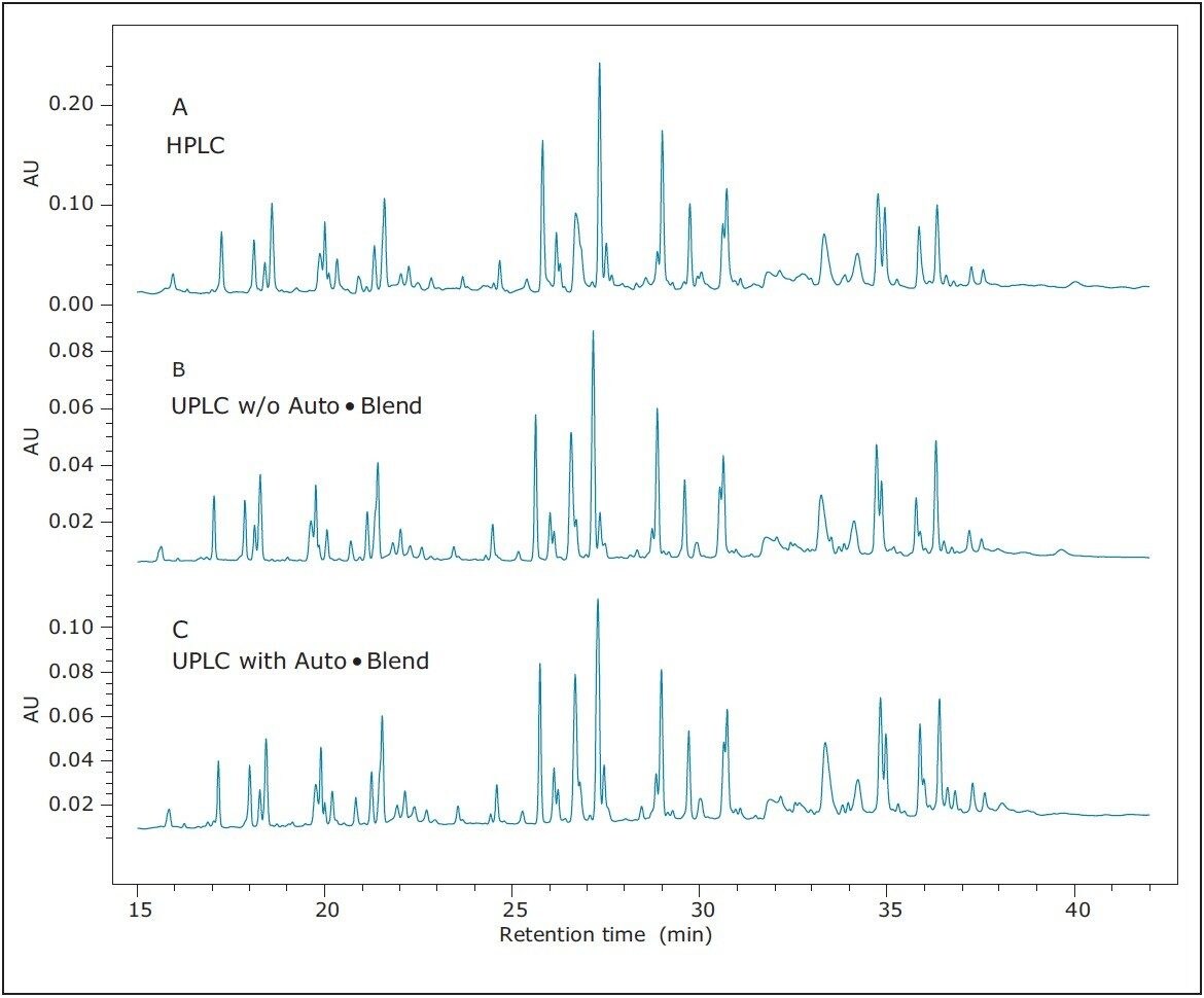
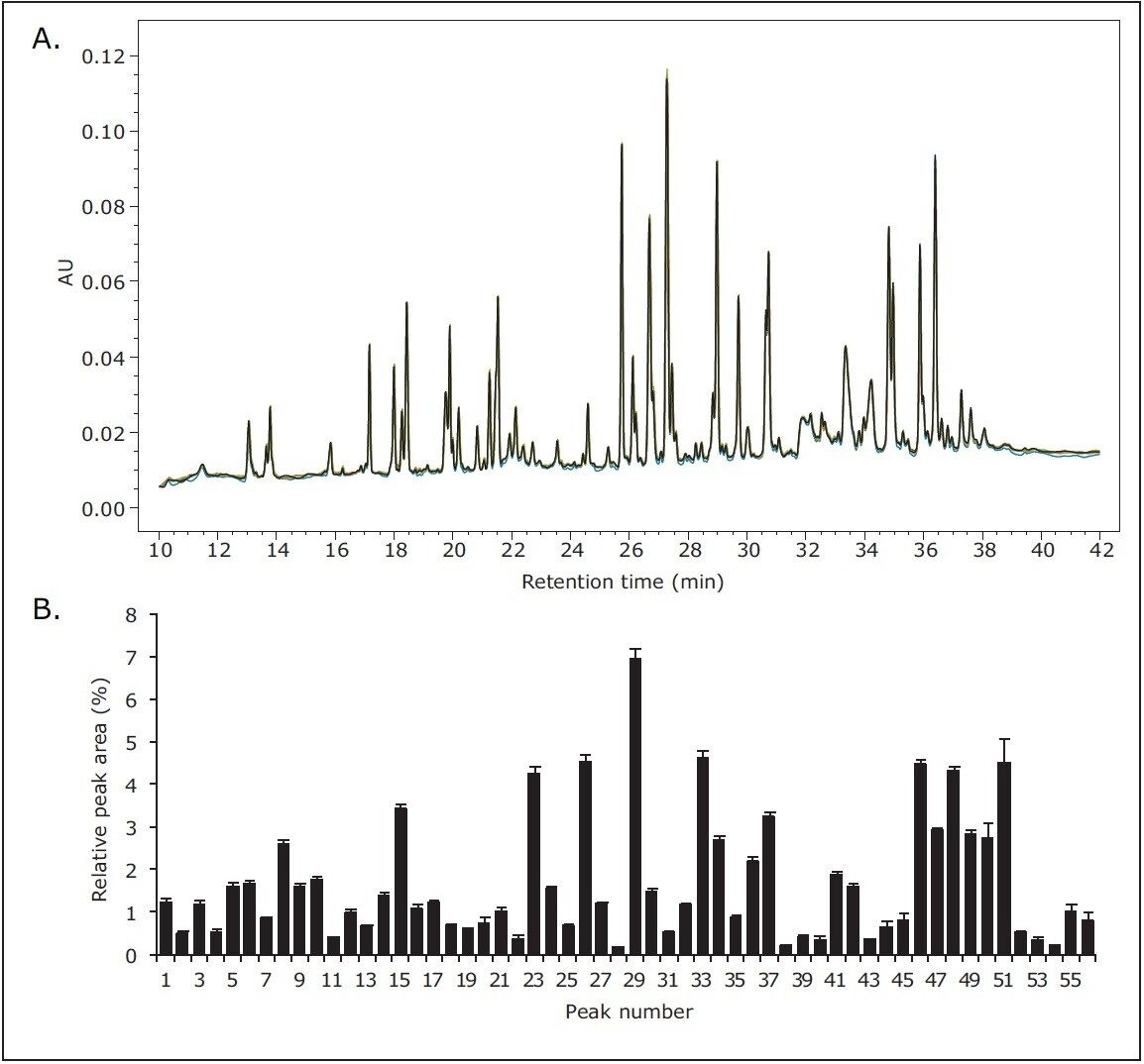
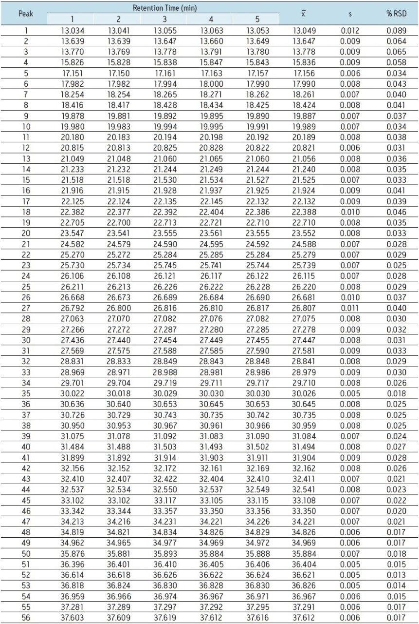
This application note demonstrates the ability of Auto•Blend Technology to control the trifluoroacetic acid (TFA) component of the mobile phase during routine peptide mapping analyses, thereby producing chromatograms of equivalent performance to HPLC-acquired chromatograms with conventionally modified mobile phases.
TFA is commonly used as a modifier in peptide mapping methods with optical detection because it provides peak shape and chromatographic resolution benefits. Concentrations of TFA are typically low in most applications, accounting for 0.02% to 0.20% of the final mobile phase. Subtle changes in the modifier concentration can have profound effects on peptide retention time, resolution, and elution order, causing concern over chromatographic reproducibility and the occurrence of out-of-specification results. Such issues ultimately affect productivity due to time-consuming resolution of QC issues as opposed to moving product to the marketplace.
The reason for this classical approach using TFA in peptide mapping chromatography has been to modify the two mobile phases, normally water and acetonitrile, with a pre-determined amount of TFA. Here, we demonstrate the benefits of allocating TFA to an independent solvent line using Auto•Blend to manage its contribution to the solvent composition throughout gradient delivery. The result is not only consistency in chromatographic performance but a significant benefit in terms of chromatographic reproducibility with minimal solvent preparatory requirements. Auto•Blend Technology in peptide mapping enables QC labs to spend less time in the prep labs, instead focusing on driving productivity.