• 应用纪要

REIMS Workflow for the Direct Analysis of Cell Pellets with Minimal Sample Preparation

REIMS Workflow for the Direct Analysis of Cell Pellets with Minimal Sample Preparation

  • Emrys Jones
  • Michael Batey
  • Waters Corporation

This is an Application Brief and does not contain a detailed Experimental section.

Abstract

Using a Waters Xevo G2-XS QTof with a REIMS source in conjunction with Progenesis QI, obtain metabolic differences between cell populations directly from cell pellets in their sample tubes, with minimal sample preparation.

Benefits

REIMS – a quick, simple, and powerful method of analyzing cell samples with minimal sample preparation.

Introduction

Rapid evaporative ionization mass spectrometry (REIMS) is an ambient mass spectrometry technique that allows materials to be sampled at remote locations from the instrument. Using radio-frequency probes, partially insulating material – such as biological tissue – is rapidly heated, leading to evaporation of the fluids present. In biological material, this evaporation causes disruption of the cells in the sampled volume, generating an aerosol that contains a large amount of molecular information. The aerosol is drawn through a length of flexible tubing and introduced to the REIMS source.

A venturi pump introduces the aerosol orthogonally to the inlet capillary of the mass spectrometer source. This allows only the smaller particulate matter to be drawn into the source, and excludes potentially problematic larger clusters due to their excessive momentum. Upon entering the source, the remaining material is directed towards a heated impactor surface where molecular ions are released to be analyzed and detected. 

Figure 1. The REIMS source and sampling device, illustrated here with the monopolar knife option. 

Results and Discussion

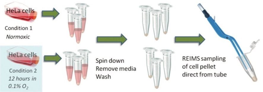
HeLa cells were grown in Minimum Essential Medium (MEM) in T75 flasks. After 24 hours of growth, half of the culture flasks were placed in a hypoxia chamber at 0.1% O2 overnight, while the other half remained under standard conditions. On the following day, the cells were suspended in 1 mL freezing media and placed into vials, resulting in approximately 2 million cells in each vial. The samples were then stored at -80 °C until analyzed.

Prior to analysis, the cells were thawed and centrifuged to allow the freezing media to be removed. 150 mM ammonium acetate was added to wash the cells. Further centrifugation was performed in order to create a cell pellet. Removal of the washing solution completed the sample preparation steps. 

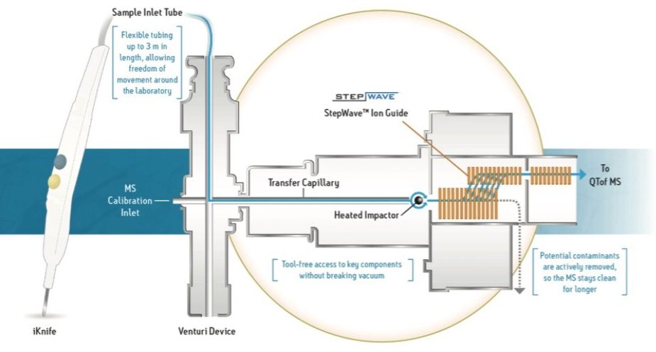
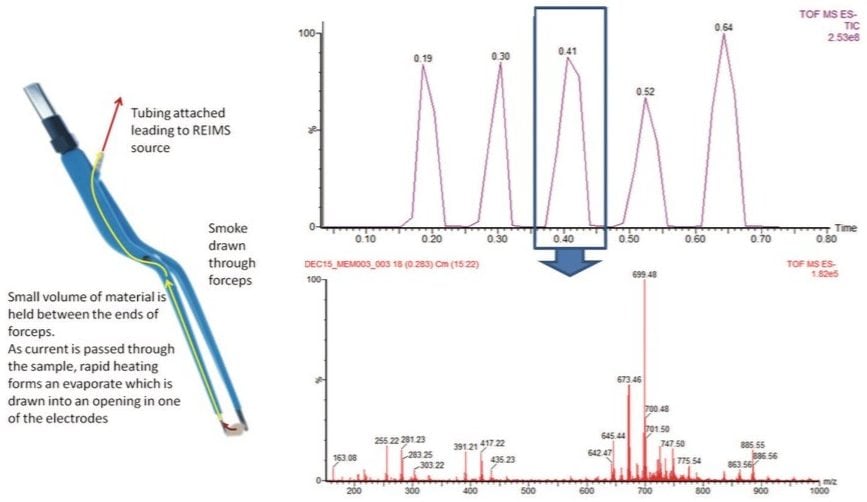
The REIMS analysis uses bipolar forceps which allow the cell pellet to be analysed in situ within the sample tube, as shown in Figure 2. A small amount of material is picked up between the two electrodes. Then, using a footswitch, the generator is activated – passing current through the cell matter. Resistive heating leads to the evaporation of the cellular cytoplasm and the disruption of the cells. The aerosol that is formed is drawn by suction through the body of the forceps and then through a length of tubing into the venturi device, to be introduced orthogonally to the inlet of the REIMS source (Figure 3). 

Figure 2. Workflow for cell pellet analysis with the bipolar forceps with REIMS. There are minimal sample preparation steps required to go from a cell suspension to obtaining mass spectrometry information from the cell population.

With this instantaneous sampling method, from initiating the current to collecting the mass spectrum, multiple data points can be collected in a matter of seconds. In Figure 3, five replicate measurements can be observed from one sample in less than one minute. The spectra obtained are rich in molecular information, especially in the lipid mass range of the spectrum. The five sampling events in the chromatogram demonstrate a high degree of intra- and inter-sample reproducibility. Figure 4 shows the spectra of five replicate sampling events from three different sample tubes of the same cell type. Once normalized to account for different sampling volumes, the CVs of most of the peaks detected are in the 10% range.

Figure 3. An illustration of the bipolar sampling device alongside a representative sample acquisition where five measurements can be made in less than a minute. The spectrum shown is of a single sampling event and demonstrates the wealth of information provided.
Figure 4. Reproducibility analysis using the bipolar REIMS technology: Five spectra from three sample tubes of the same cell type. The CVs are typically below 10% for all peaks within the data set. 

Having a rapid and reproducible analysis technique allows for easy comparison of the metabolic profiles of different cell types or cells subjected to different conditions. To complement the ease of analysis, Progenesis QI provides a streamlined data analysis solution. Using the Progenesis Bridge Application, MassLynx Software data – with multiple sampling events in each raw data – are separated into individual spectra that can then be imported into Progenesis QI as data points. Figure 5 shows the principal component analysis (PCA) results of the hypoxic versus normoxic cell comparison. Within the software, differences between the samples can be demonstrated and identifications suggested for the molecules contributing to the differentiation. From the initial analysis of three samples, each of the hypoxic and normoxic cell pellets lipid species – at increased and decreased levels in the hypoxic cells – are identified.

Figure 5. Results of analysis of REIMS data in Progenesis QI. The replicates of the two biological conditions clearly separate from each other on the first principal component. The subsequent plots show lipid species that are at elevated levels in the hypoxic sample, elevated in the normoxic sample and at equal intensity in both samples. 

Conclusion

Rapid evaporative ionization mass spectrometry (REIMS) provides a quick, simple, and powerful method of analyzing cell samples with a breadth of possible applications.

No sample preparation, other than washing and spinning the cells into a pellet, is required and the measurement can be made directly from the sample tube.

The data can be easily imported in Progenesis QI where a range of analysis and identification tools can be applied to maximize the information obtained from the experiment.

Thank you to Fiona Henderson, Dr. Adam McMahon, and Prof. Kaye Williams of the University of Manchester for their help in culturing and analyzing the cells.

720005774, August 2016

返回顶部 返回顶部