• Application Note

Parallel MALDI-Tof Post Source Decay Analysis of a Complex Protein Mixture

Parallel MALDI-Tof Post Source Decay Analysis of a Complex Protein Mixture

  • Emmanuelle Claude
  • Marten F. Snel
  • Daniel Kenny
  • Jeffery M. Brown
  • Therese McKenna
  • James I. Langridge
  • Waters Corporation

Abstract

  • A new MALDI post source decay (PSD) multiplexed (MX) technique is presented
  • This technique allows for the acquisition of PSD spectra from all precursor ions simultaneously, thus removing the serial nature of a conventional MALDI PSD experiment
  • Coupling this approach to an off-line HPLC separation, with deposition of the eluent onto a MALDI target plate, allows for comprehensive investigation of complex peptide mixtures
  • The new parallel MALDI PSD approach is compared and contrasted to MALDI MS/MS on a Q-Tof Mass Spectrometer

Introduction

Liquid chromatography followed by mass spectrometry is the accepted approach for analyzing complex protein mixtures. Electrospray ionization has usually been the preferred technique as it allows comprehensive on-line LC-MS/MS to be performed. However, in recent years there has been a growing interest in the use of MALDI ionization for analyzing complex tryptic digest mixtures that have been separated by off-line HPLC. This approach provides complementary information to that obtained by ESI, while deposition onto a MALDI target allows samples to be archived for further analysis.1

In this application note two types of MALDI mass spectrometer were used to analyze complex tryptic peptide mixtures, after HPLC separation. The instruments used in this study were a Waters Micromass Q-Tof Ultima MALDI Mass Spectrometer and a Waters Micromass MALDI micro MX Mass Spectrometer. The MALDI micro MX is equipped with a new MALDI MS/MS mode of operation, which provides structural information. This technique is a novel form of post source decay (PSD) analysis, termed PSD MX. In traditional PSD the selection of precursor ions with a timed electrostatic iongate is required. In the PSD MX approach, the ion gate is not required as fragment ions from all of the precursor ions are acquired simultaneously. A deconvolution algorithm has been developed to match precursor ions with fragment ions.2

In order to establish proof of principle prior to the analysis of a more complex sample, a mixture of 15 standard proteins was digested using trypsin and separated by reverse phase HPLC. A more complex mixture was produced from an Escherichia coli (E. coli) cytosolic cell fraction, which was also digested and reverse phase HPLC separated. The eluent from these two separations was spotted directly onto Waters standard 96-well MALDI target plates, using a modified Waters 2700 MS spotting robot. Matrix solution was co-deposited with the HPLC eluent. In total, 48 MALDI target wells from the 15-protein mixture and 24 MALDI target wells from the E. coli lysate were analyzed with the Waters MALDI micro MX and Q-Tof Ultima MALDI mass spectrometers.

Experimental

Mass Spectrometry

MALDI micro MX

The MALDI micro MX incorporates a MALDI source and an axial ToF mass analyzer with a reflectron detector for recording both MS and PSD MX data. Data were acquired in positive ion mode using automated software control. In the MS mode, a digest of alcohol dehydrogenase (ADH) was used to generate a multi-point external calibration. An external lock mass correction was applied using ACTH (18-39 clip). In PSD MX mode, data were calibrated using PSD fragments generated from ACTH (18-39 clip).

Q-Tof Ultima MALDI

The Q-Tof Ultima MALDI instrument combines a MALDI source with a quadrupole as the first mass analyzer, which allows the selection of the peptide precursor ions. These precursor ions are transmitted through a hexapole gas cell where they undergo multiple low-energy collisions to induce collision decomposition using defined collision energies. The product ions produced in the hexapole collision cell are then mass measured using an orthogonal acceleration time-of-flight mass spectrometer.

Data were acquired in positive ion mode using the Data Directed Analysis (DDA) Software. Selected ions were fragmented by collision induced dissociation (CID) to provide MS/MS product ion data. Polyethylene Glycol (PEG) was used as an external multi-point calibration and subsequently an external lock mass using [Glu]1-Fibrinopeptide b was applied.

Sample Preparation

15-Protein mixture

  • An equimolar mixture of 15 protein tryptic digests was produced containing: Phosphorylase B (Rabbit), Serum albumin (Bovine), Enolase (Yeast), Lysozyme (Hen egg white), Lactoperoxidase (Bovine), Glucose oxidase (A. niger), Carbonic anhydrase (Bovine), Catalase (Bovine), Alcohol dehydrogenase (Yeast), Cytochrome C (Horse), Fetuin (Bovine), Myoglobin (Horse), Alpha Lactalbumin (Bovine), Beta casein (Bovine), Alpha casein (Bovine).
  • Each protein was dissolved separately to 100 μg/mL in 0.1 M NH4HCO3 subsequently reduced with DTT (4 h at room temp) and alkylated with iodoacetamide (4 h room temp). Each protein was then digested with sequencing grade trypsin (Promega, Madison, WI) for 4 h at 37 °C. The solutions were then diluted and mixed to give a final concentration of 100 fmol/μL.

Escherichia coli cytosolic fraction

  • An aliquot of 500 μL of K12 derived E. coli, cytosolic fraction, was diluted in 0.1% RapiGest SF in 25 mM ammonium bicarbonate, 0.5 M DTT, 1 M CaCl2 and digested by adding 400 μg of Promega sequencing grade trypsin in resuspension buffer. After digestion at 37 °C for 14 hours, a second aliquot of trypsin was added and the digestion continued for an additional 4 hours. Finally, the sample was centrifuged at 12,000 g for 10 min. The resulting supernatant was collected and diluted five times in an aqueous solution containing 0.1% TFA, to a final protein concentration of 1 μg/μL.

MALDI LC Conditions

The complex tryptic digest mixtures (2 μL of the E. coli sample and 5 μL of the 15-protein mixture respectively) were injected and separated by reverse phase chromatography on a Waters CapLC System using a Waters NanoEase C18 column 150 x 0.075 mm and the following conditions:

  • Flow rate was 8 μL/min, split to 200 nL/min pre-column
  • Mobile phase A (ACN/0.1% TFA in H20, 5:95)
  • Mobile phase B (ACN/0.1% TFA in H20, 95:5)
  • Gradient initial conditions 5% B changed to 40% B over 90 minutes

The entire flow from the HPLC column was spotted onto a 96-well MALDI target plate using the 2700 MS spotting device. A solvent delay of 12 minutes was applied before the beginning of the sample deposition. Each MALDI target position had 30 seconds of HPLC eluent applied, equivalent to 100 nL. In parallel, alpha-cyano-4-hydroxycinnamic acid was co-deposited at a rate of 1.8 μL/min.

PSD MX Technology

  • As precursor ions are not selected using a traditional ion gate, those which decompose by PSD give rise to a mixture of fragment ions that are detected simultaneously. These fragments are matched to their associated precursor ion by acquiring two time-of-flight spectra at slightly different reflectron voltages. Since fragment ions have a unique combination of mass and kinetic energy related to the mass of the precursor, it’s possible to determine the precursor of each fragment by measuring the shift in time-of-flight of the same fragment ion in the two spectra.
  • A spectrum acquired at the same reflectron voltage as conventional PSD is called the major spectrum. A spectrum acquired approximately 4% lower is called the minor spectrum.
  • In both traditional and parallel PSD (PSD MX) experiments, small low energy fragment ions do not penetrate as deeply into the back of the reflectron as their respective precursors and consequently are not as well focused. This limitation is overcome by acquiring several major spectra (commonly known as segments) at reduced reflectron voltages. Stitching together the focused regions of each segment give rise to a single fragment ion spectrum.

Results and Discussion

15-protein Mix Results

Multiplexed PSD and MALDI Q-Tof data were acquired from a total of 48 wells containing the HPLC fractionated mixture of 15 standard proteins. The resulting fragment ion data were combined into a single peaklist file for each instrument and searched against the SwissProt protein database, using MASCOT (Matrix Science). Figure 1 shows a comparison of the MS/MS data obtained from a tryptic peptide of Alpha lactalbumin using PSD MX and the Q-Tof Ultima MALDI. Table 1 displays a summary of the database search results obtained.

Table 1. Summary of identified proteins from 15-protein mixture.
  • Using the MALDI micro MX, twelve of the fifteen proteins were correctly identified (where a correct identification is defined as an ion score >37).
  • A total of four proteins obtained ion scores greater than the identification level, but were incorrect (false positive identifications).
  • With the Q-Tof Ultima MALDI, all 15 proteins were correctly identified.
  • The results for the two analyses of the 15 proteins mixture are summarized in Table 1.

E. coli Results

Figure 2 displays MS spectra from six consecutive HPLC fractions of the E. coli protein mixture. These spectra clearly show the efficiency of the HPLC separation since there is little overlap of peptides between adjacent spots.

PSD MX analysis of each fraction and subsequent data processing produced “clean” PSD (fragmentation) spectra for each precursor ion present in the sample. These deconvoluted PSD MX spectra showed similar fragmentation to CID spectra from MALDI-MS/MS analysis on the same sample with the Q-Tof Ultima MALDI, shown in Figures 3 and 4.

Results from the analysis for both the MALDI micro MX and the Q-Tof Ultima MALDI are summarized in Table 2. Entries in red illustrate a positive identification with a confident score. Those proteins with a yellow background show better identification using MALDI micro MX, while those with a grey background show better identification using the Q-Tof Ultima MALDI. Those identifications shown in black text illustrate a tentative identification, with a low confidence level.

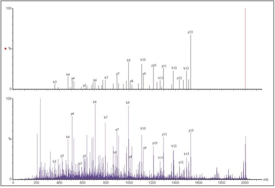
For each instrument, the fragment ion data from all 24 spots were combined into a single peaklist file, which was database-searched using MASCOT (Matrix Science).

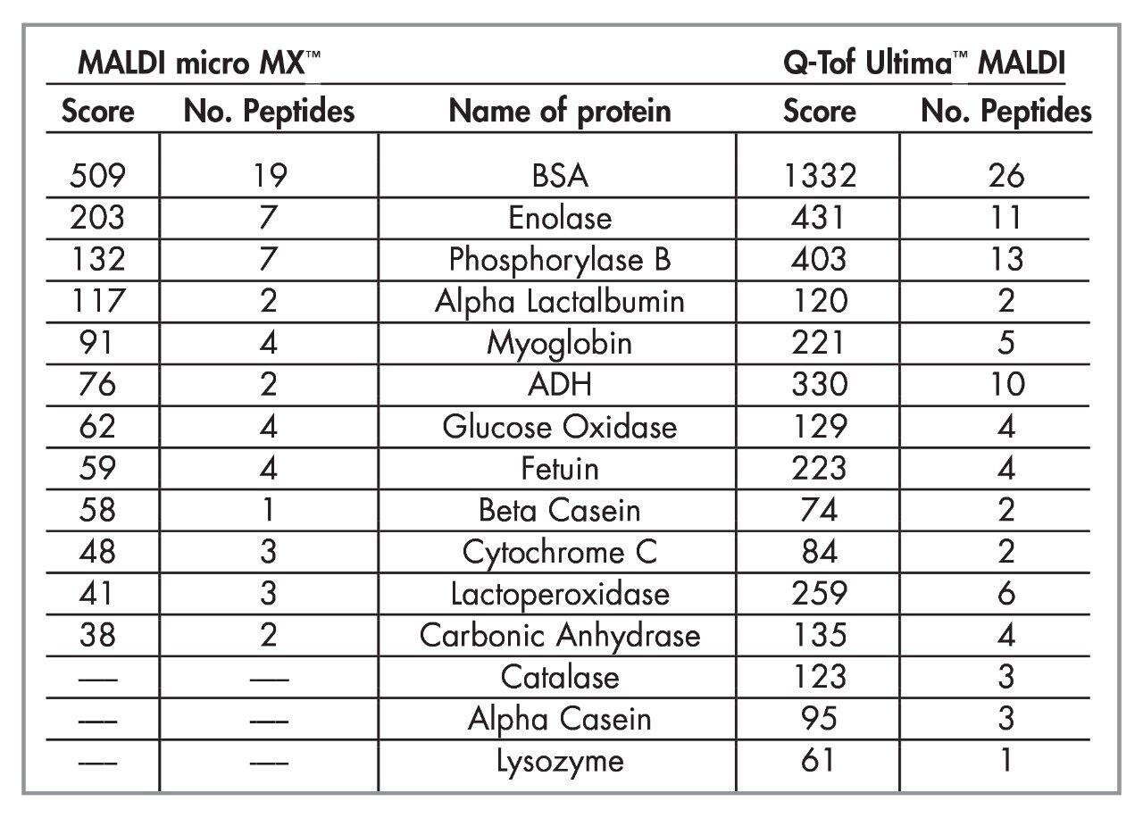
  • The combination of the MALDI micro MX and the Q-Tof Ultima MALDI resulted in the identification of 14 proteins with significant confidence levels from the databank search.
  • Of these 14 proteins, five proteins were common to the two techniques, while three proteins were only identified using the MALDI micro MX. An additional six proteins were only identified using the Q-Tof Ultima MALDI.
  • An additional seven proteins were identified using PSD MX with a score below the positive confidence level. Of these, two were confirmed by subsequent analysis on the Q-Tof Ultima MALDI.
  • In addition, four proteins were identified using Q-Tof Ultima MALDI with a score below the positive confidence level, while analysis of these by MALDI micro MX was able to confirm one of these identifications with a high score.

These results indicate that the use of MALDI on a Q-Tof geometry and parallel PSD on an axial instrument can been seen as complementary techniques. The use of both techniques provides significantly better coverage of the proteins present than either technique alone.

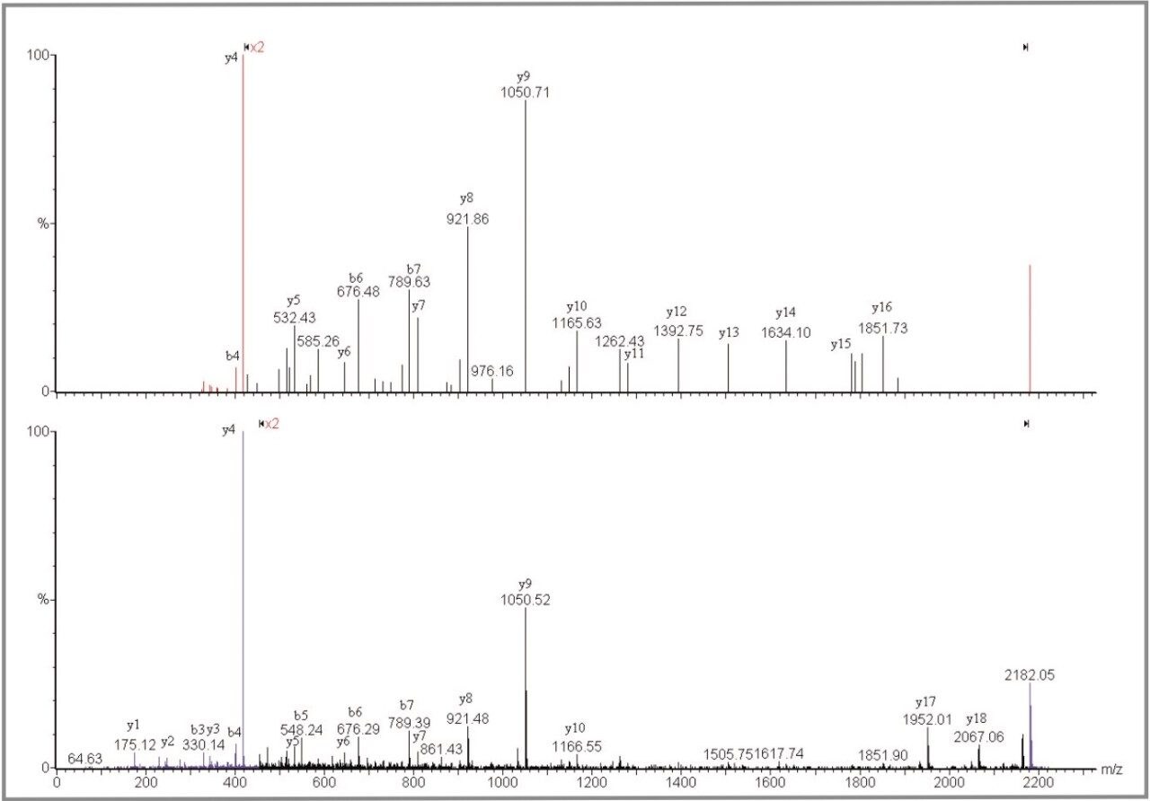
The advantage of the Q-Tof Ultima MALDI instrument is the high mass measurement accuracy. The average mass accuracy on the 21 peptides confidently identified is 2.8 ppm. The mass measurement accuracy of the Q-Tof Ultima MALDI provides a considerable improvement in the specificity of the overall experiment. This enables significantly increased confidence to be obtained from the database search, or de novo amino acid sequence to be obtained in certain cases. As can be seen in this dataset, the MS/MS fragmentation pattern from one peptide is sufficient to positively identify a protein.

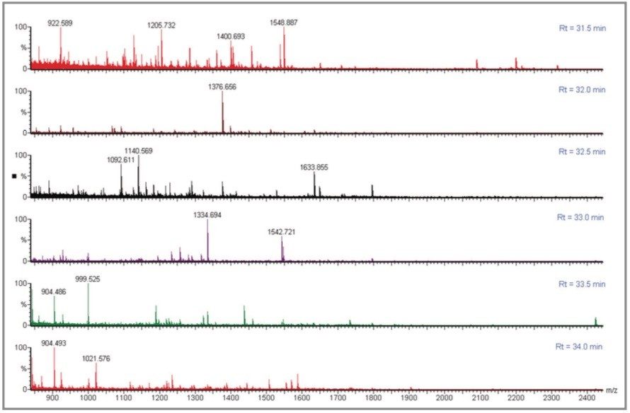
In comparison, the strength of the PSD MX acquisition is the parallel nature of the experiment. This has significant benefits in terms of automating the acquisition, and greatly simplifies the experiment. For example, the protein OSMC_ECOLI has not been identified with the Q-Tof Ultima MALDI but was confidently identified in the PSD MX experiment. The precursor ions at m/z 1140.6 Da and 2155.1 Da were not automatically selected using the Q-Tof Ultima MALDI as other more intense ions were present in the spectrum, and were selected for MS/MS. In this case the parallel nature of the PSD MX experiment provides complementary information, from multiple precursor and fragment ions, which in turn enhances the specificity.

Conclusion

  • A novel parallel approach to PSD is presented
  • This allows for the acquisition of PSD spectra from all precursor ions simultaneously, thus removing the serial nature of a conventional MALDI PSD experiment
  • Time and sample are not wasted acquiring data on peptides that do not fragment well by PSD
  • This approach simplifies the automated acquisition of PSD spectra and reduces the time required to generate data from multiple precursors
  • Off-line nanoscale HPLC separation in combination with deposition onto a MALDI target plate provides significant benefits by reducing the sample complexity
  • Data generated using this approach compares favorably to that obtained from a MALDI source equipped Q-Tof instrument
  • The two MALDI techniques are complementary and the combination provides more information than either technique alone

References

  1. J.P.C. Vissers, R.K. Blackburn, M.A. Moseley, A Novel Interface for Variable Flow Nanoscale LC-MS/MS for Improved Proteome Coverage, J. Am. Soc. Mass Spectrom. 2002, 13(7), 760–771.
  2. D. Kenny, J. Brown, M. Snel, Technical Note: Multiplexed Post Source Decay (PSD MX): A Novel Technique Explained

Featured Products

720001044, December 2004

Back To Top Back To Top