Haemophilus influenzae is a major pathogen responsible for a wide variety of human infections. This virulent bacterium causes serious diseases such as meningitis, epiglottitis, bacteremia, cellulitis, acute otitis media, and sinusitis. The physiology of this pleomorphic, Gram-negative coccobacillus bacterium has been well characterized over recent years and it was the first genome of a free-living organism to be completely sequenced (Fleischmann et al., 1995).
The cell walls of H. influenzae contain lipooligosaccharide, which resembles the lipopolysaccharide of Gram-negative bacilli but has shorter side chains (hence the designation oligosaccharide rather than polysaccharide). H. influenzae does not make toxins or other extracellular products that account for their ability to produce infection. The mechanism of pathogenesis of H. influenzae infections is not completely understood. However, the presence of the type b polysaccharide capsule is a major factor in virulence.
Encapsulated organisms can penetrate the epithelium of the nasopharynx and invade blood capillaries directly.
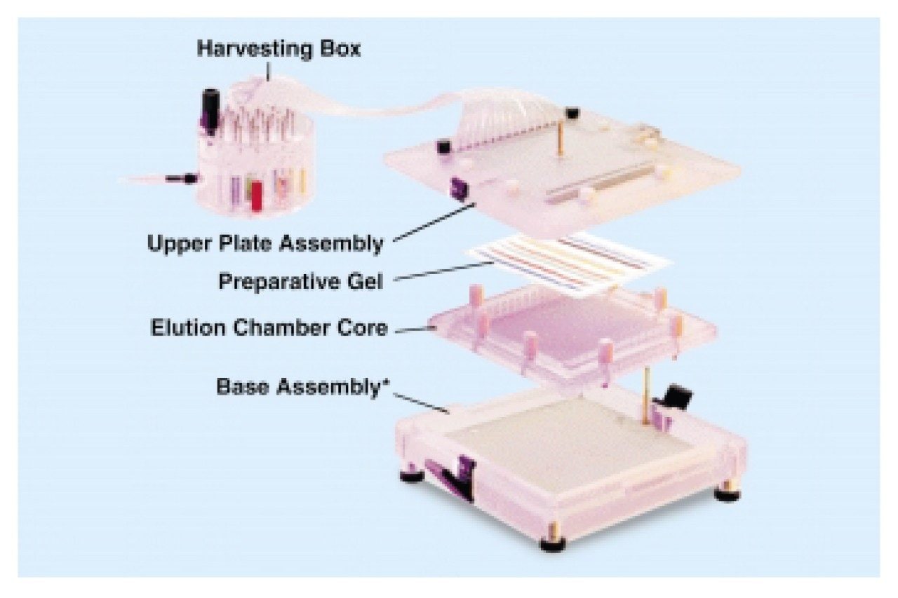
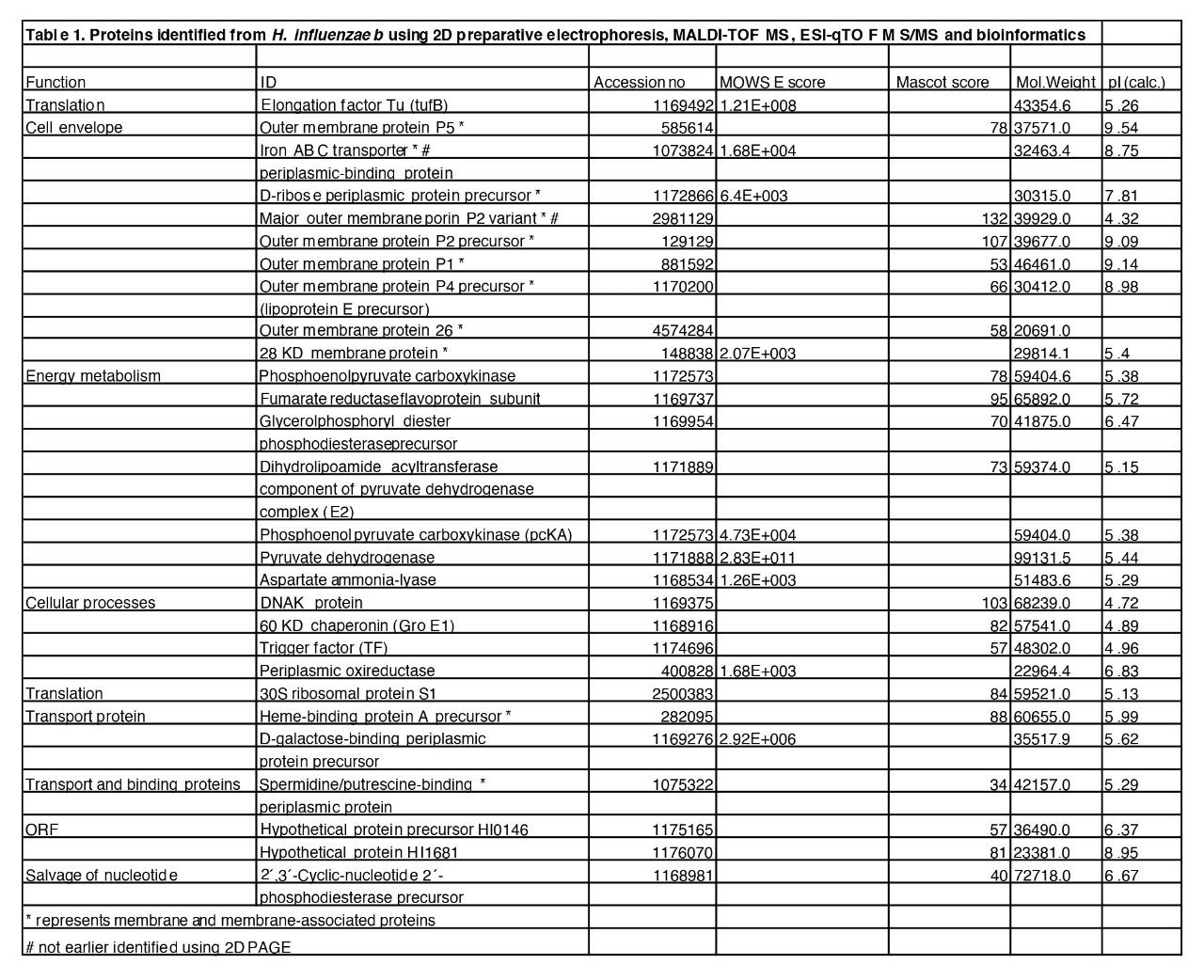
The bacterium use pili and fibrils during the process of attachment and colonisation of the host. These cell surface macro structures contain proteins that may be potential vaccine candidates. However, identification of membrane associated proteins is difficult using traditional proteomics techniques such as 2D PAGE. This is because the membrane proteins contain hydrophobic transmembrane domains that span the phosopholipid membrane. The result of these hydrophobic domains is that the protein is difficult to solubilise and will not remain in solution during the isoelectric focusing on an IPG strip.
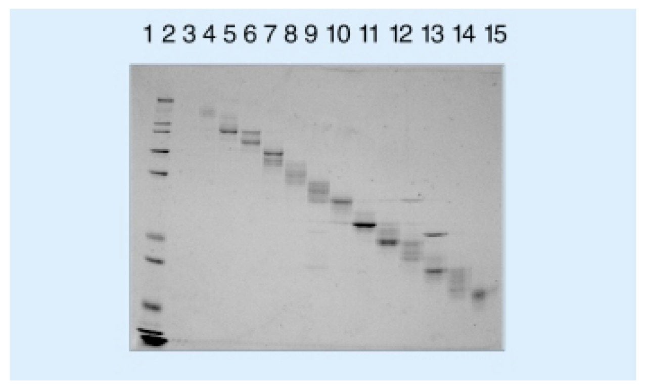
In this paper we have used two dimensional preparative electrophoresis to separate proteins in an extract from H. influenzae b.