There is increasing interest in the analysis of spatial distribution of small molecules in tissues for drug discovery, disease diagnosis, or biomarker discovery. Localization of the dosed drug and its metabolites are critical information for understanding the mechanism of target-organ toxicity.
Matrix-assisted laser desorption ionization (MALDI) is a sensitive solid-sampling and soft-ionization technique with extensive applications for the analysis of both large and small molecules. The MALDI mass spectrometry (MS) signal can be easily obtained directly from tissue sections.1 The resulting three-dimensional image becomes very useful for the investigation of localization of dosed drug and its metabolites in tissue.
MALDI imaging provides an alternative to whole-body autoradiography in that there is no need to use a radiolabel to trace the drug and its possible metabolites throughout the organ of interest. This means that substantial savings are made, and, as a consequence, scientists can conduct an imaging experiment in targeted organs earlier on in the discovery process without the need of having a synthesized, radiolabeled new chemical entity.
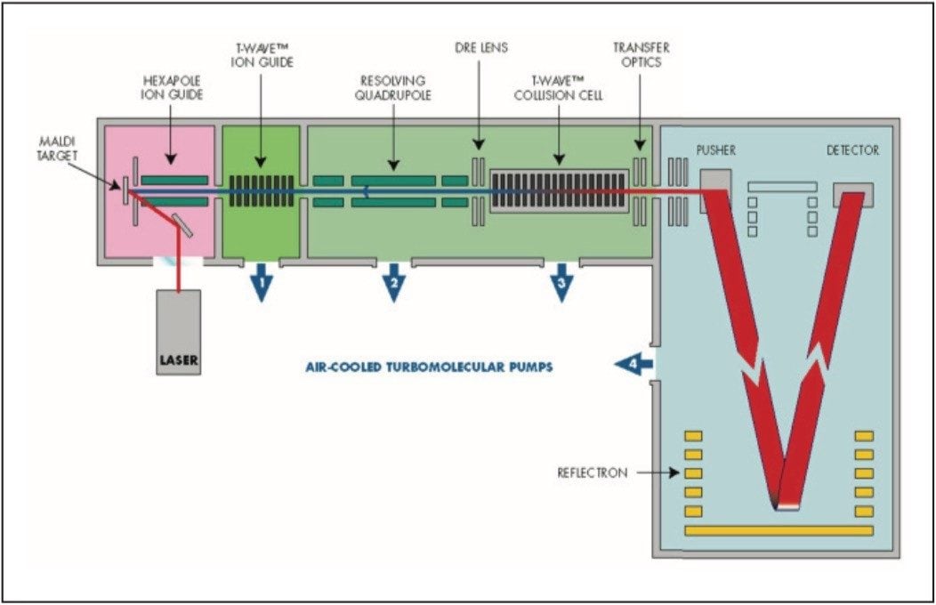
Combining MALDI with quadrupole time-of-flight (TOF) MS, utilizing the Waters MALDI Q-Tof Premier Mass Spectrometer, offers excellent sensitivity and selectivity for these tissue imaging experiments.
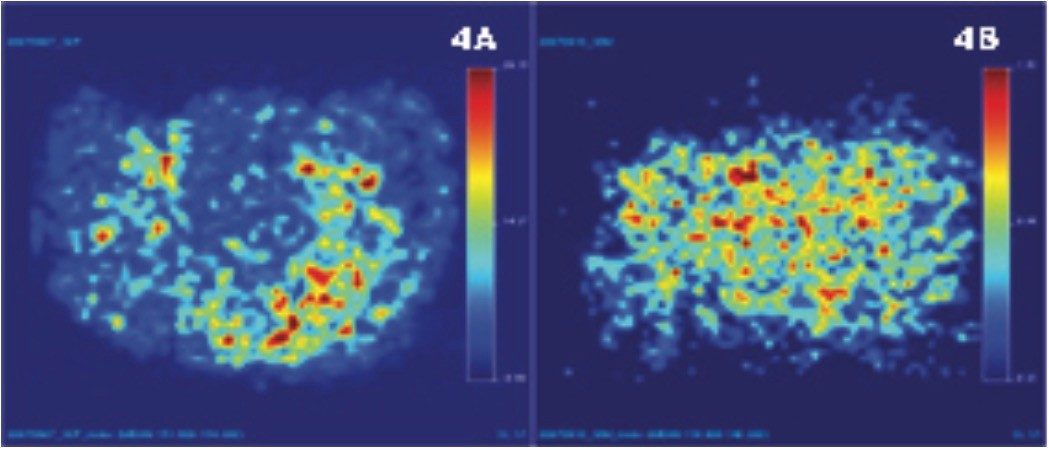
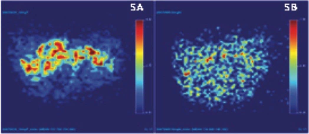
In this application note, we use diazepam as an example to demonstrate the utility of MALDI TOF MS in this application area. After diazepam was intravenously administered to rats, a study using sliced rat brain tissues was performed. Results obtained showed clear localization of the parent drug and its metabolite.
Therefore, MALDI TOF MS proved sensitive, specific, and highly amenable to the image analysis of traditional small molecule drug candidates directly in tissues.