Automated RapiFluor-MS Labeled Glycan Sample Preparation for Disulfide-Rich Glycoprotein
Abstract
N-glycans are routinely monitored for the quality of biotherapeutics because they affect the safety and efficacy of many therapeutic proteins. Current glycan analysis is developed with the well-studied glycans from monoclonal antibodies (mAb). Here, we present a complementary method that provides a reproducible profile of N-glycans released from Human Chorionic Gonadotrophin (hCG), a highly complex glycoprotein with multiple disulfide bonds. The automated reduced protein protocol is suitable for commercial as well as research environments.
Benefits
- Rapid automated protocol for 32 samples in 1 hour
- Reproducible glycan profiles from complex and disulfide-rich glycoprotein
- QC-friendly protocol suitable for commercial environment and research
Introduction
N-glycans Analysis is Challenging, yet Required for Routine Analysis
Many therapeutic glycoproteins are composed of N-glycans that play critical roles in the folding, conformation, and stability of the proteins.1 N-glycans are sugars that are covalently attached to the glycoproteins at the nitrogen atom of an asparagine (Asn) side chain.1 Unlike a chemical synthesis, the biosynthesis of N-glycans is a concerted effort involving multiple enzymes at various stages and consequently, yields several diverse structures of N-glycans – high-mannose, hybrid, and complex.1 Owing to such structural heterogeneity, the analysis of glycans is filled with many technical challenges.2
Highly sensitive to the manufacturing conditions, glycans also affect the efficacy and clinical safety of biotherapeutics.2 Therefore, drug developers and manufacturers have to monitor, characterize, and identify the glycans released from glycoproteins to ensure the quality of biotherapeutics.2 In addition, N-glycosylated proteins are analyzed as biomarkers for several diseases such as testicular cancer3 and ectopic pregnancy.4 These glycoproteins associated with diseases are increasingly becoming more complex in their structures and heterogeneity.4 One approach to facilitate the release of the N-glycans is to reduce the disulfide bridges in the glycoprotein in order to help unfold the glycoprotein, exposing not easily accessible N-glycans for release with PNGase F. Human chorionic gonadotropin (hCG), is a good candidate for this approach due to its highly complex structure. hCG is mainly known for its role in ectopic pregnancy and consists of complex type N-glycans and multiple disulfide bridges as shown in Figure 1.4
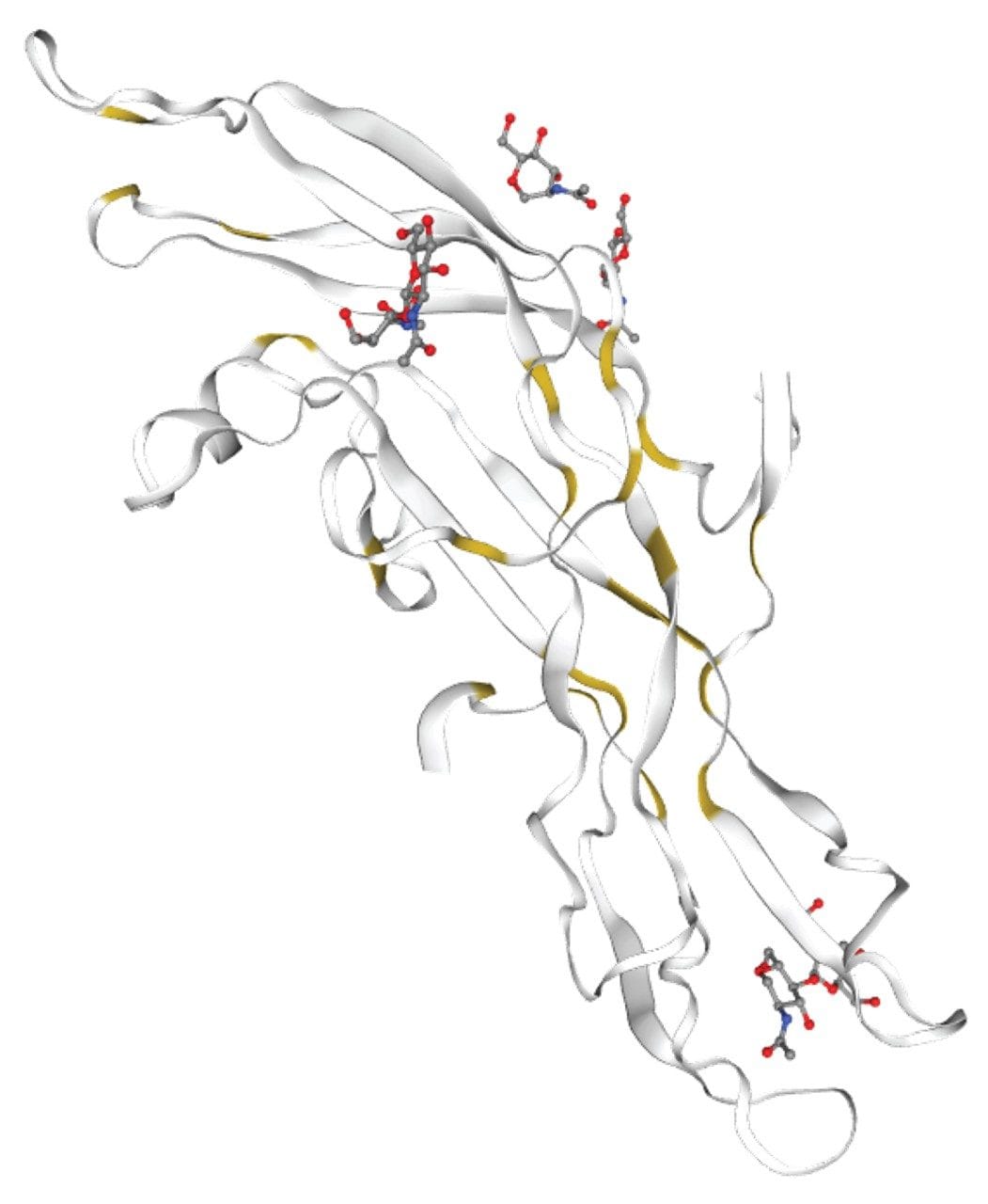 Figure 1. Structure of Human Chorionic Gonadotropin (hCG) with N-glycans (red) and disulfide bonds (yellow). Figure generated from the website (swissmodel.expasy.org).
Initially, for disulfide-rich glycoproteins, the GlycoWorks RapiFluor-MS procedures5,6 outlined the use of Tris (2-carboxyethyl)phosphine (TCEP) as a reductant. While the TCEP reduction is compatible with the HILIC separation of RFMS-labeled glycans, the by-products of the TCEP reduction have been shown to interfere with the reversed-phase mixed-mode (RP/AX) separation of RFMS-labeled glycans. As a result, a procedure has been developed using an optimized amount of glycoprotein and dithiothreitol (DTT) as a reductant that does not cause significant interferences for N-glycans with the RP/AX separation.
GlycoWorks RapiFluor-MS Reducing Protocol Provides a Rapid Streamlined Solution to Complex N-glycan Analysis
The new reducing protocol is similar to the original GlycoWorks RapiFluor-MS (non-reducing) protocol on the Andrew+ Pipetting Robot,7 except two reagents are introduced – Dithiothreitol (DTT) and ammonium acetate. DTT is used to reduce the disulfide bonds that can hinder the effectiveness of PNGase F enzyme for some N-glycans. Ammonium acetate is used as a quenching solution to minimize an interference peak when using RP/AX columns such as ACQUITY Premier Glycan BEH C18 AX Column (p/n: 186009758). Ammonium acetate is optional if HILIC columns are used. Figure 2 shows an overview of the reducing protocol which includes 3 steps: 1) reduce and deglycosylate, 2) label and quench, 3) clean up.
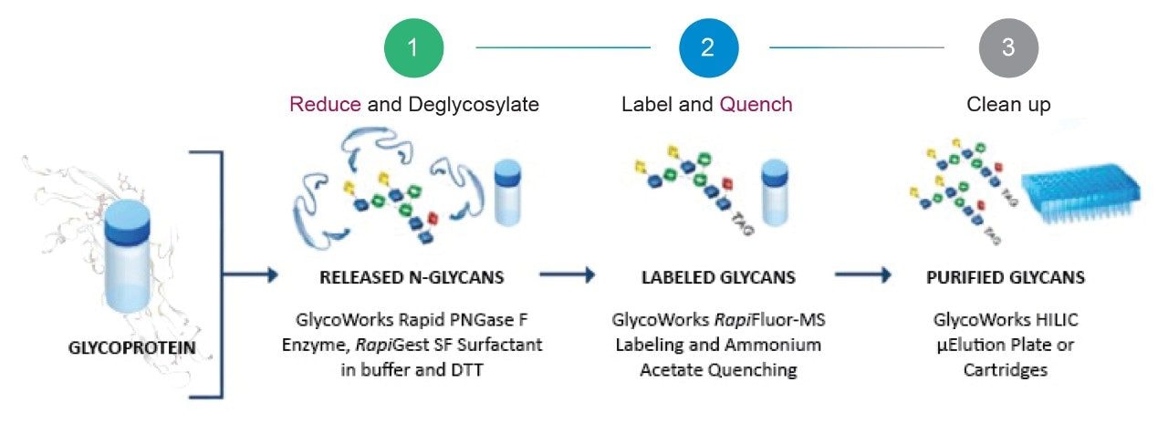 Figure 2. Workflow for the rapid preparation of complex N-glycans using the GlycoWorks RapiFluor-MS reducing protocol. Quenching with ammonium acetate is optional for HILIC separation.
Experimental
Sample Description
Manual sample preparations can be found in these protocols- 720006992EN8 and 720006991EN.9 Adjustments to these protocols were made to accommodate the labware used and maintain a high recovery of glycans for the automation protocol. Three solutions were prepared first. The Intact mAb Mass Check Standard (p/n: 186006552) (1 mg/vial) was reconstituted in 830 µL 18.2 MΩ water to obtain a 1.2 mg/mL mAb solution. RapiGest Surfactant (p/n: 186008090) 3% with 2 µg/µL DTT denaturing buffer was prepared by first dissolving 3 mg of RapiGest in 60 µL of the GlycoWorks Rapid Buffer (p/n: 186008939) and adding the 40 µL of the 5 µg/µL DTT solution (water) into the surfactant mixture. Rapid PNGase F enzyme (30 µL) (p/n: 186008939) was diluted with 220 µL of water. Deglycosylation was begun by dispensing the 10 µL of glycoprotein at 1.2 mg/mL in a 96 well plate, followed by the additions of 10 µL of 3% (w/v) RapiGest with 2 µg/µL DTT denaturing buffer. After heat denaturation at 90 °C for 3 minutes, the solution was cooled to ambient temperature and 10 µL of PNGase F solution was added. Deglycosylation was continued at 50 °C for 5 minutes. The RapiFluor-MS (RFMS) (p/n: 186008091) solution was obtained by adding 110 µL of anhydrous DMF to the vial of 9 mg of RFMS reagent and aspirating to mix. The resulting deglycosylated solution was labeled by adding 15 µL of RFMS solution. The labeling reaction was allowed to continue for 5 minutes at ambient temperature. Ammonium acetate solution (10 µL, 0.5 M) was added to quench the reaction for 5 minutes at ambient temperature.
To prepare for the purification of N-linked glycans, the GlycoWorks HILIC µElution Plate (p/n: 186002780) was first conditioned with 200 µL of water, and equilibrated with 200 µL of 85% acetonitrile. The resulting N-glycan mixture was diluted with 495 µL acetonitrile and loaded on the plate. Samples are washed with four volumes of 300 µL of 1% formic acid, 90% acetonitrile to remove the impurities and excess labeling reagent. Lastly, the N-glycans were eluted using three 30 µL volumes of elution buffer (p/n: 186007992) and then diluted with 310 µL sample diluent (p/n: 186007992) to obtain purified released glycans.
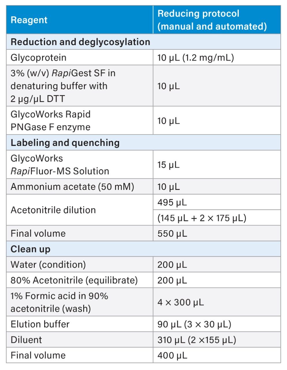 Table 1. Volumes for the manual and automated reducing protocols used for this application note. Refer to experimental session above for more details on sample dilutions.
LC Conditions
|
LC system: |
ACQUITY UPLC H-Class PLUS Bio System |
|
Detection: |
FLR detector 265 nm (ex) 425 nm (em)10 |
|
Sample collection: |
twin.tec PCR Plate 96, skirted, green, Eppendorf, (p/n: 951020443) |
|
Column: |
ACQUITY UPLC Glycan BEH Amide Column, 1.7 µm, 2.1 mm x 150 mm, 130 Å p/n: 186004742 |
|
Column temp.: |
60 °C |
|
Sample temp.: |
10 °C |
|
Injection volume: |
10 µL |
|
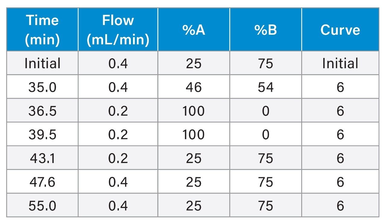
Mobile phase A: |
50 mM Ammonium formate, pH 4.4 (LCMS grade, p/n: 186007081) |
|
Mobile phase B: |
Acetonitrile |
Gradient
Data Management
|
Chromatography software: |
Empower 3 |
Results and Discussion
Automation of N-glycans Analysis via GlycoWorks RapiFluor-MS Reducing Protocol
Automation of routine N-glycan analysis is highly suitable for QC environment as well as general research for high consistency and reproducibility. The automated reducing GlycoWorks RapiFluor-MS protocol can deliver a highly reproducible profile of complex N-glycans from disulfide rich glycoprotein. Steps for the automated reducing protocol are shown in the flow diagram below.
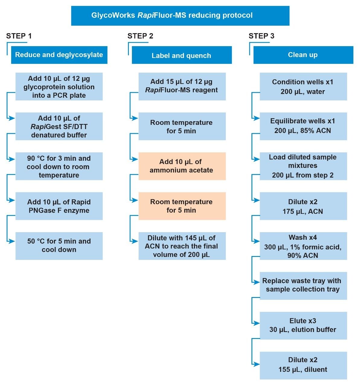 Figure 3. Flow diagram of the automated GlycoWorks RapiFluor-MS Reducing protocol. Quenching step with ammonium acetate, as highlighted in orange, is optional for HILIC separation.
For this procedure, the Andrew+ Pipetting Robot was placed inside a chemical hood (with the dimension of 72W x 33.5D; 13.8 sq. ft.; Model: HBBV6, Lab Crafters. Inc.). Two configurations of dominos are shown in Figure 4 and 5. The configuration in Figure 5 uses 3 rows of dominos and will execute the reducing protocol in its entirety. Users electing to use a smaller chemical hood, such as the one described above, would need to use the 2-step configuration shown in Figure 5 (A) and (B), which uses only 2 rows of dominos. “Step A” protocol will execute reduction and deglycosylation, labeling, and quenching, and “Step B” will execute the purification.
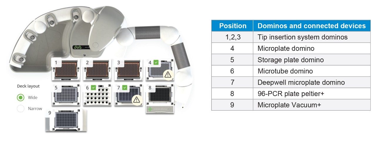 Figure 4. Andrew+ domino configuration for the rapid automated GlycoWorks RapiFluor-MS reducing 32-sample protocol. Configuration is for 3 rows of dominos. The execution time for 32 samples is 1 hour.
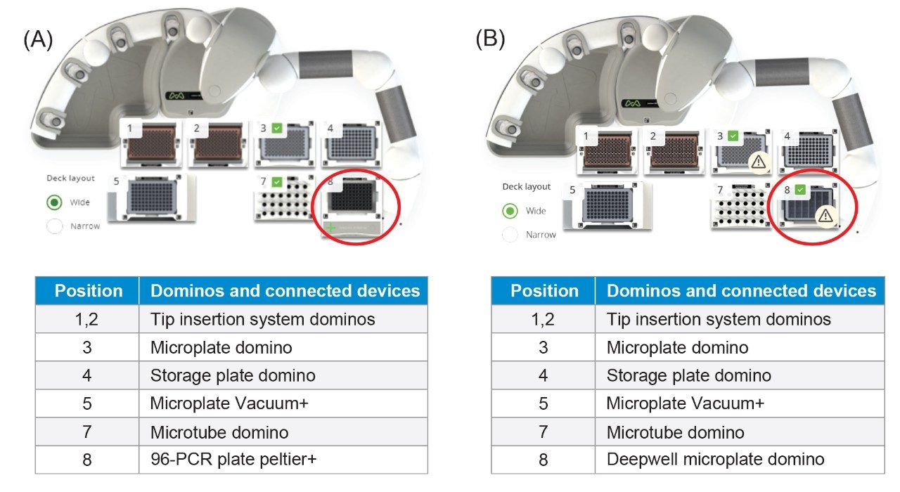 Figure 5. Andrew+ domino configuration for the rapid automated GlycoWorks RapiFluor-MS reducing 32-sample protocol (A) step 1 (B) step 2. These configurations use 2 rows of dominos to accommodate a smaller chemical hood. The total execution time for 32 samples is 1 hour 10 minutes.
Rapid Automated GlycoWorks RapiFluor-MS Reducing Protocol Can Be Used for Profiling the N-glycans From Disulfide-rich Glycoproteins
Using the reducing protocol, Andrew+ provided consistent glycan profiles for monoclonal antibody (mAb). Initial optimization was performed using the Intact mAb Mass Check Standard (p/n: 186006552) to act as a control for the RFMS-labeling portion of the procedure since N-glycan release from the mAb is effective without disulfide bond reduction. The reducing protocol gave comparable recovery of released N-glycans. Figure 6 shows the comparison of the chromatograms obtained from the reducing protocol (Figure 6A) and the non-reducing protocol (Figure 6B). No differences in the total peak areas were observed for all four major glycoforms released from the glycoprotein.
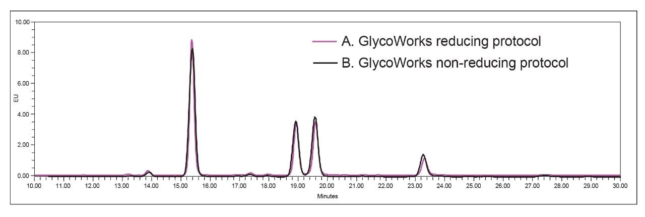 Figure 6. Glycans released from Intact mAb Mass Check standard using (A) GlycoWorks RapiFluor-MS reducing protocol and (B) GlycoWorks RapiFluor-MS non-reducing protocol. The amount of released glycans were similar for both protocols.
Figure 7 compares the relative areas and the total peak areas of selected (FA2, FA2G1a, FA2G1b, and FA2G2) released glycans from mAb of the manual (6 samples) and automated executions (6, 16, or 32 samples) of the protocol. For each of these sample preparations, 6 of the prepared samples were analyzed by UPLC. Relative areas obtained for the 4 selected glycans observed for both manual and automated experiments are comparable (%RSD of FA2 ≤5% as shown in Figure 7A. The largest percent deviation (5% for FA2) was observed between the 6-sample manual (FA2 = 42%) and 32-sample automated (FA2 = 44%) procedures.
The total peak areas were consistent for the manual and automated glycan sample preparations, as can be seen in Figure 7B. The largest percent difference in average peak area (14%) was observed for FA2 when comparing the 16 and 32 sample automated experiments. A critical aspect of glycan sample preparation is to avoid failed preparations. In these experiments, the largest individual recovery difference between two sample preparations as represented by the relative peak area range [100 x (max-min)/mean] was observed for FA2, 6 sample manual procedure and was 21%.
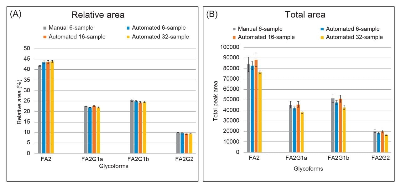 Figure 7. Comparison of glycan profiles released from the manual execution (6 samples) and the Andrew+ Liquid Handling Robot (6, 16, or 32 samples). Intact mAb Mass Check Standard (p/n: 186006552) was used for these experiments.
Human Chorionic Gonadotropin (hCG) was also selected to test the reducing protocol because of its highly complex structure and multiple disulfide bridges. The structure of hCG consists of a dimer representing two highly glycosylated subunits, and four N-linked glycans along with multiple disulfide bonds.4 The N-glycans attached are mostly of the complex-type.4
Reducing protocol enabled the release of N-glycans that are hard to access when using the non-reducing protocol. Figure 8 shows the difference in glycan profiles between the two protocols. The reduction of DTT during denaturation allowed more glycans to be accessible by the PNGase F enzyme for deglycosylation. The increased amount of glycans released was evident in peaks marked with (*) in the reducing protocol (black trace). The three peaks labeled with (*) were higher for the reducing protocol (black trace) than the ones labeled with (*) in pink trace.
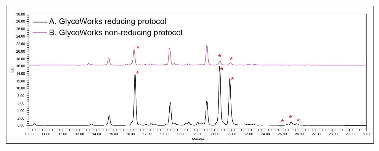 Figure 8. Glycans released from Human Chorionic Gonadotropin (hCG) using (A) GlycoWorks RapiFluor-MS reducing protocol and (B) GlycoWorks RapiFluor-MS non-reducing protocol.
Reducing protocol increased the amount of released glycans on the peaks marked with (*). Using the rapid automated reducing protocol, Andrew+ provided consistent glycan profiles released from hCG. Figure 9 compares the relative areas and the total peak areas of the selected released glycans from hCG of the manual (6 samples) and automated executions (6, 16, or 32 samples) of the protocol. For each of these sample preparations, 6 of the prepared samples were analyzed by UPLC. Relative areas obtained for the 9 selected glycans observed for both manual and automated experiments are comparable (%RSD of peak 5 and 6 ≤5%, as shown in Figure 9A. The largest percent deviation (7% for peak 5) was observed between the 6-sample automated (peak 5 = 28%) and 16-sample automated (peak 5 = 30%) procedures.
The total peak areas were consistent for the manual and automated glycan sample preparations, as can be seen in Figure 8B. The largest percent difference in average peak area (12%) was observed for peak 5 and peak 6 when comparing the 6-sample manual and the 6-sample automated experiments. The largest individual recovery difference between two sample preparations as represented by the relative peak area range [100 x (max-min)/mean] was observed for peak 5, 6 sample manual procedure and was 25%.
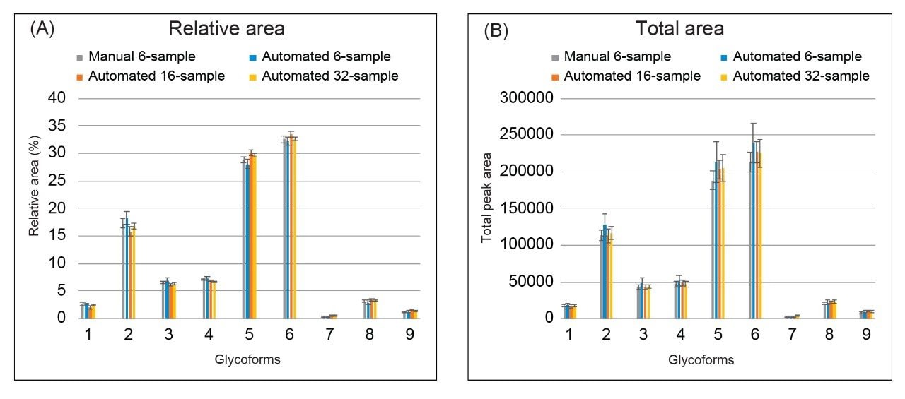 Figure 9. Comparison of N-glycan profiles released from the manual execution (n=6) and the Andrew+ Liquid Handling Robot (n=6, 16, and 32). Human Chorionic Gonadotropin (hCG) was used for these experiments.
Conclusion
Highly sensitive to the manufacturing conditions, glycans are routinely monitored and characterized as a critical quality attribute (CQA) of protein biotherapeutics. Automation of routine N-glycan analysis is invaluable for general research as well as process and product development. The automated sample preparation method follows the GlycoWorks RapiFluor-MS Released N-glycan method with adjustments outlined in this paper that include the reduction of glycoprotein using dithiothreitol (DTT). The relative abundance as well as recovery of released N-glycans for samples prepared on Andrew+ using the reducing protocol were comparable to those observed with manually executed experiments. In summary, we present an automated and complementary method that can provide reproducible N-glycan profiles from disulfide rich glycoproteins with complex glycans such as hCG, as demonstrated here.
Considerations
1. Recommended starting glycoprotein quantity
- This automated reducing protocol is designed to produce optimal results from 12 μg of glycoprotein. Samples with a concentration of 1.5 mg/mL can still potentially produce appropriate results provided that the volume of the labeling reagent (RFMS) is adjusted accordingly. Significant changes to the optimal glycoprotein quantity, i.e. <10 μg, can affect the Rapid PNGase F enzyme to substrate ratio as well as the molar excess of RFMS labeling reagent, which will potentially result in undesirable labeling artifacts or low recovery of glycoforms.
2. Regarding the RapiFluor-MS Reagent:
- RapiFluor-MS is a highly reactive, primary/secondary amine labeling reagent. It hydrolyzes in water with a half-life on the order of 10–100 seconds. It is, therefore, important that the reagent be dissolved in the provided anhydrous DMF, a non-nucleophilic, polar aprotic solvent. Reagent solution can be used across the span of a day if care has been taken to limit exposing the solution to atmospheric moisture.
- The reducing protocol uses twice the amount of RFMS, compared to the non-reducing protocol because the reducing agent, DTT, is nucleophilic and interferes with the labeling reaction by consuming RFMS. As a result, 16 reduced protein glycan labeling preparations can be generated from a 24-sample kit (Andrew+ 24 Sample GlycoWorks Application, p/n: 176003349) and 32 preparations can be generated from a 48-sample kit (Andrew+ 96 Sample GlycoWorks Application, p/n: 176003350 or Andrew+ 96HT Sample GlycoWorks Application, p/n: 176003351).
3. One step or two steps reducing protocol
- If the Andrew+ Robot is inside a chemical hood, users can choose which automated protocol to use, depending on the size of the chemical hood. The protocol with 3 rows of dominos carries out the experiments in its entirety. The 2-step protocol (STEP A and STEP B) with 2 rows of domino configurations are included to accommodate a smaller size of chemical hood (with the dimension of 72Wx33.5D; 13.8 sq. ft.; Model: HBBV6, Lab Crafters. Inc.).
4. Microplate Gripper
- Using a gripper (p/n: 186009776) is recommended for the reducing protocol carried out inside a chemical hood. The microplate gripper moves the 96-well reaction plate between a domino and the Peltier+ before and after the heat denaturing steps. This transfer will help to obtain comparable glycan recovery between the manual and automated protocols.
References
- Varki, A.; Cummings, R. D.; Esko, J. D.; Stanley, P.; Hart, G. W.; Aebi, M.; Darvill, A. G.; Kinoshita,T.; Packer, N. H.; Prestegard, J. H.; Schnaar, R. L.; Seeberger, P. H. Essentials of Glycobiology, Third Edition. 2017. Cold Spring Harbor (NY): Cold Spring Harbor Laboratory Press; 2015–2017. doi: 10.1101/glycobiology.3e.00.
- Zhang, P.; Woen, S.; Wang, T.; Liau, B.; Zhao, S.; Chen, C.; Yang, Y.; Song, Z.; Wormald, M. R.; Yu, C.; Rudd, P. M. Challenges of Glycosylation Analysis and Control: An Integrated Approach to Producing Optimal and Consistent Therapeutic Drugs. Drug Discovery Today. 2016, 21 (5), 740–765.
- Hires, M.; Jane, E.; Mego, M.; Chovanec, M.; Kasak, P.; Tkac, J. Glycan Analysis as Biomarkers for Testicular Cancer. Diagnostics (Basel, Switzerland), 2019, 9(4), 156.
- Biskup, K.; Blanchard, V.; Castillo-Binder, P.; Alexander, H.; Engeland, K.; Schug, S. (2020). N- and O-Glycosylation Patterns and Functional Testing of CGB7 versus CGB3/5/8 Variants of the Human Chorionic Gonadotropin (hCG) Beta Subunit. Glycoconjugate Journal, 2020, 37(5), 599–610.
- Lauber, M. A.; Yu, Y.-Q.; Brousmiche, D. W.; Hua, Z.; Koza, S. M.; Magnelli, P.; Guthrie, E.; Taron, C. H.; Fountain, K. J. Rapid Preparation of Released N-glycans for HILIC Analysis Using a Labeling Reagent That Facilitates Sensitive Fluorescence and ESI-MS Detection. Anal. Chem. 2015, 87 (10), 5401–5409.
- Koza, S. M.; McCall, S. A.; Lauber, M. A.; Chambers, E. E. Quality Control and Automation Friendly GlycoWorks RapiFluor-MS N-glycan Sample Preparation. Waters Application Note, 720005506EN, Revised 2020.
- Reed, C. E., Koza, S., Calciano, S. Automated Medium and High-Throughput GlycoWork RapiFluor-MS Preparations on the Andrew+ Pipetting Robot. Waters Application Brief, 720007008EN, 2020.
- GlycoWorks RapiFluor-MS Quick Start Protocol – 24 Sample. Waters Brochure, 720006992EN, 2021.
- GlycoWorks RapiFluor-MS Quick Start Protocol – 96 Sample. Waters Brochure, 720006991EN, 2020.
- Lauber, M. A.; Brousmiche, D. W.; Hua, Z.; Koza, S. M.; Guthrie, E.; Magnelli, P.; Taron, C. H.; Fountain, K. J. Rapid Preparation of Released N-glycans for HILIC Analysis Using a Novel Fluorescence and MS-Active Labeling Reagent. Waters Application Note, 720005275EN, 2015.
720007402, October 2021