Use of the Andrew+™ Robot and OneLab™ Automated Liquid Handling Platform for the Fungitell® Glucan Detection Assay
This is an Application Brief and does not contain a detailed Experimental section.
Abstract
To demonstrate that Andrew+ and OneLab can be configured to execute the Fungitell® glucan detection assay.
Introduction
The Fungitell assay is a 96 well microplate based Limulus Amebocyte Lysate (LAL) based colorimetric assay that measures (1→3)-β-D-glucan (BDG), a major cell-wall component of various medically important fungi.1 Fungitell is FDA cleared and CE marked for the detection of BDG in the serum of patients with symptoms of, or medical conditions predisposing the patient to invasive fungal infection.2,3 This LAL based assay is an extremely sensitive enzymatic cascade method capable of detecting glucan in the picogram per milliliter (pg/mL, 10-12 g/mL) range.3,4 The assay is a fairly complex process necessitating numerous pipetting steps. Standard curve preparation, sample preparation, sample arraying, and reagent dispensing are time consuming and tedious but are critical steps in these assays. Automation of the liquid handling steps used in standard curve construction and sample creation; and utilization of automation for the arraying samples and standards on the plate represents improved efficiency for these assays. The automated process liberates the analysts from repetitive time consuming operations, leading to increased productivity, better quality in analytical work, and consistency in execution. In addition the added benefit of the OneLab event log would ensure a fully auditable process simplifying any future analyses and investigations.
LAL glucan assay execution note: As mentioned above these assays detect minute quantities of glucan in the picogram per milliliter range. As indicated in the IFU all materials used for assay execution must be tested for potential interferences. Excellent lab technique and a clean environment are required to execute these assays. These results were collected with the Andrew+ in a static enclosure.
Here we present example results from the execution of the Fungitell assay with Andrew+ and OneLab utilizing the standard pipettes and dominos. Dominos are modular holders for tubes, plates, tips, reagents, and other materials that can be configured in various combinations on the deck. Figure 1 shows the physical set up of the robot and the deck with the associated dominos.
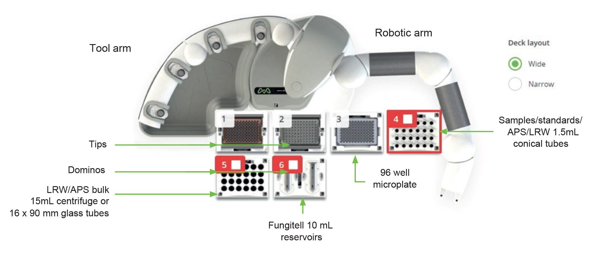 Figure 1. Andrew+ Configuration.
This is the configuration used for the Fungitell assay to produce the results described in this technical note. The electronic Bluetooth connected pipettes used were the 300 µL multichannel, 300 µL single channel pipette, and a 10 µL single channel pipette. LRW is LAL reagent water and APS is alkaline pretreatment solution.
Results and Discussion
This initial evaluation of the Andrew+ system addressed the creation of an OneLab protocol to execute the Fungitell using currently available dominos. The protocol was designed to evaluate the capability of the Andrew+ and OneLab to create a standard curve and execute sample preparation. This evaluation examined the outcomes of the robotically generated assay results for consistency in the execution of the standard curve serial dilutions and sample creation from multiple runs.
A key element of the Fungitell method is the construction of the standard curve. For each plate described herein a new standard curve was produced from a new set of dilutions. These dilutions were created as serial dilutions 300 µL to 300 µL starting with a 500 pg/mL sample. This creates a five point standard curve with concentrations of 500, 250, 125, 62.5, and 31.25 pg/mL. The standards were loaded onto the plate in duplicate as directed by the protocol. The standard curve rate data (mAbs/min) for each standard curve on each plate were analyzed by executing a linear regression producing a slope, intercept, and R-value for each of the runs. For this set of standard curve data, eight runs were executed over two days. These results are summarized in Figure 2.
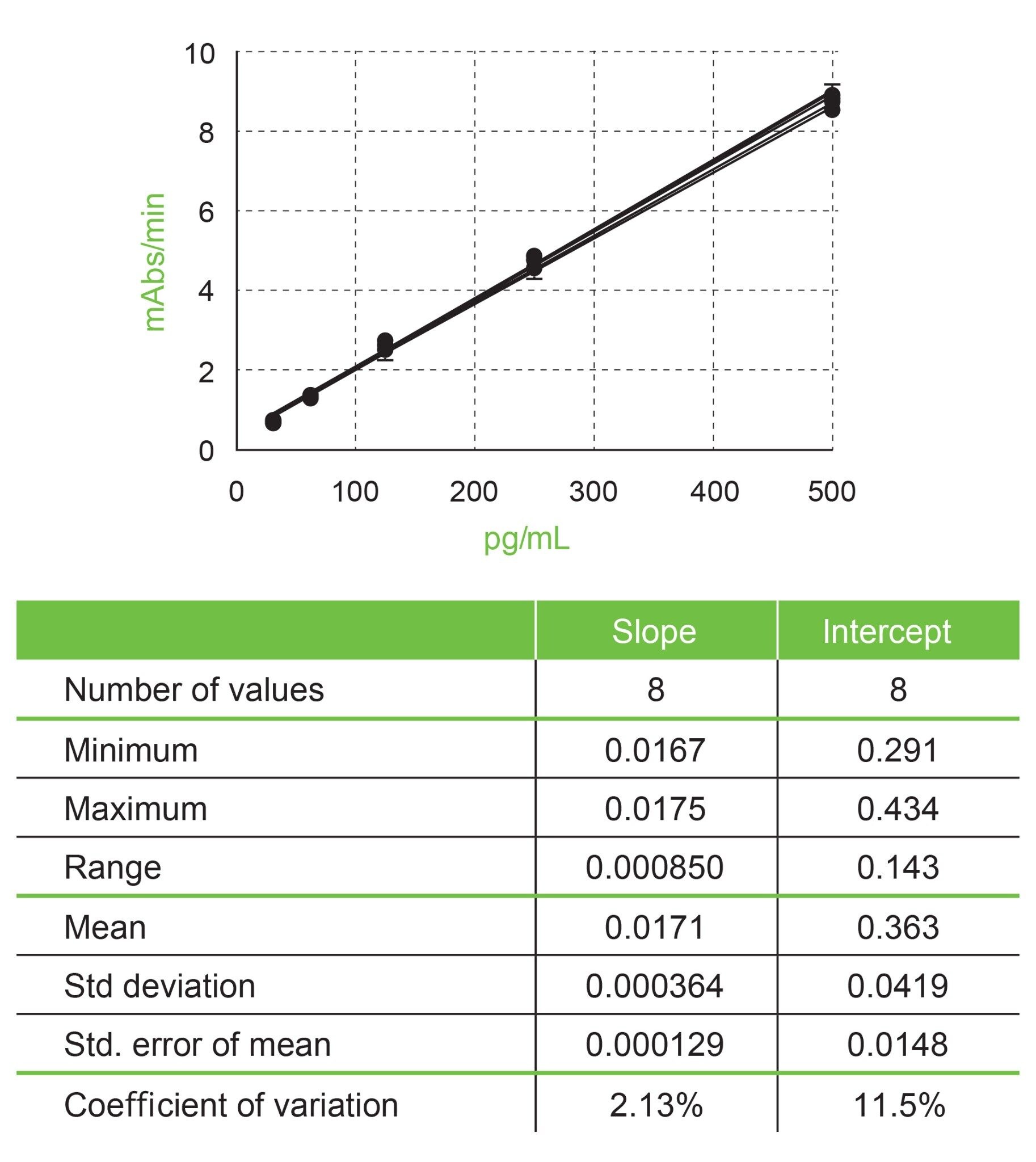 Figure 2. Standard Curves for Fungitell executed by Andrew+.
This set of data (eight runs) used the same lots of reagents and glucan standard on two different days executing the same protocol set up. The standard curve for each plate was constructed and dispensed in duplicate from a unique set of dilutions. The standards for these curves were 500, 250, 125, 62.5, and 31.25 pg/mL(See the Fungitell IFU for more explanation of these values). Each set of duplicate data were fit with a standard linear regression. Each x-axis point in this graph contains 16 data points.
The variance for slopes and intercepts across the eight runs was 2.3% and 11.5%, respectively. The R-values were all excellent with the lowest one being 0.997 (the requirement is R ≥0.980). These results are all within acceptance criteria for the Fungitell assay as indicted in the IFU.
In addition to the standard curve evaluation, various samples were tested in these experiments. The creation of the sample in the plate is a critical part of the Fungitell assay and one that can lead to error. The samples are created in the plate by adding 5 µL of sample and 20 µL of alkaline pretreatment solution to each well. The addition of 5 µL of a viscous serum sample can sometimes lead to significant variance since each addition is a unique pipetting step. In one set of experiments (Figure 3) three samples were serially diluted in a sugar solution to mimic the viscosity of serum. These three samples were made through manual dilution and tested through the typical Fungitell process.
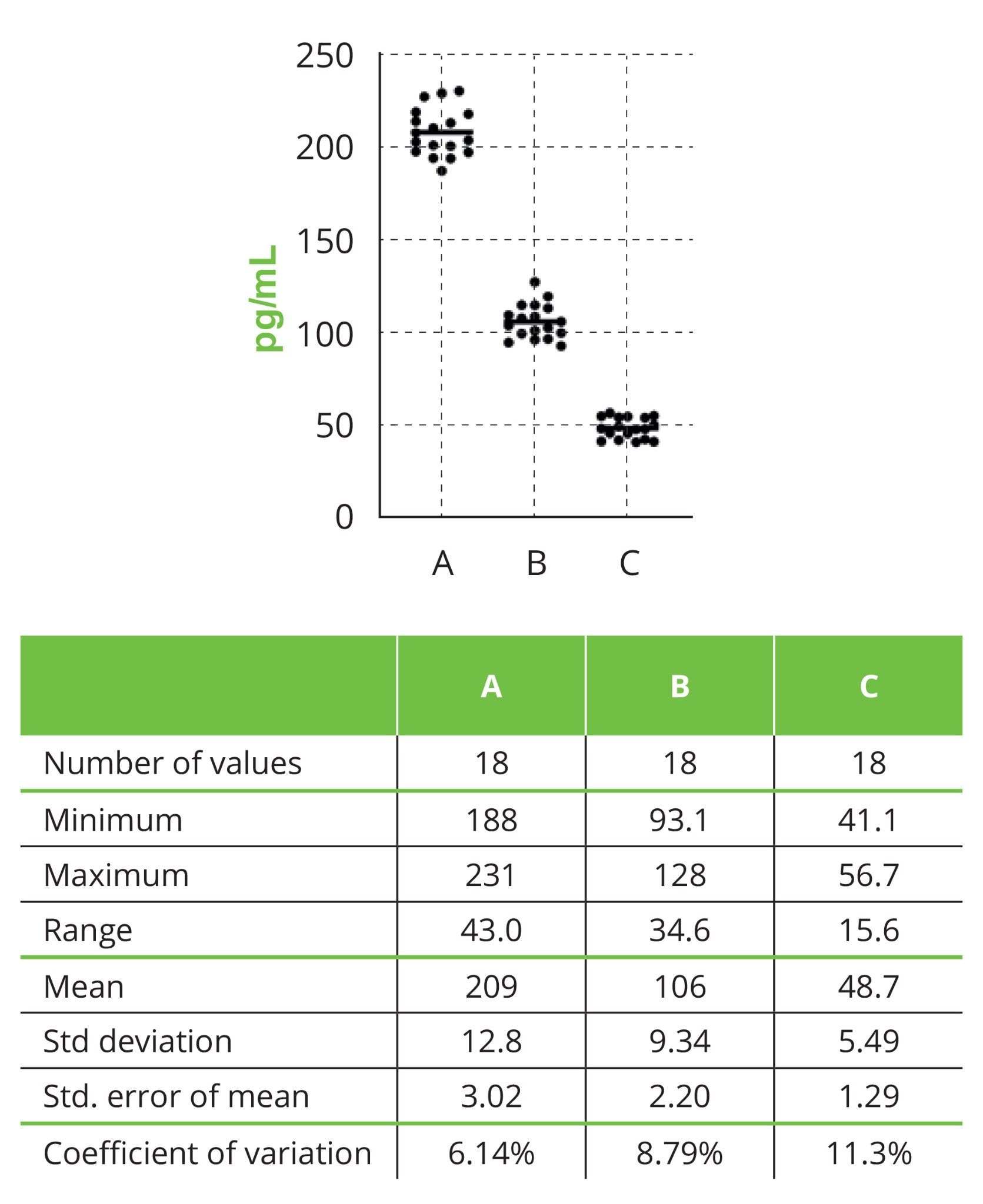 Figure 3. Sugar based sample results for Fungitell executed by Andrew+.
The scatter plot contains eighteen points per sample and the line in each set is the mean. Each sample was run on each of three plates six times and the entire set of eighteen points is included in this analysis. Each plate was a full execution complete with new standard curve creation. These experiments were executed on the same day across three plates using the same lot of reagents.
They were designed to deliver results for sample concentrations at approximately 180, 90, and 45 pg/mL, respectively. The goal was to examine the variance of the sample results across plates and at different concentrations. On each plate each sample was run six times and the eighteen results were analyzed. The variance for samples ranged from 6 to 11%. The outcome demonstrates excellent consistency for each of the samples.
In another set of experiments two serum samples were examined. These samples were created using commercially available normal off-the-clot sera spiked with glucan. The concentrations targeted were 250 to 300 pg/mL and 125 to 150 pg/mL, respectively. These two samples were tested across three plates in replicates of ten. Each serum sample was loaded a unique 5 µL pipetting step as described above. The data from across the three plates were examined and the results are summarized in Figure 4.
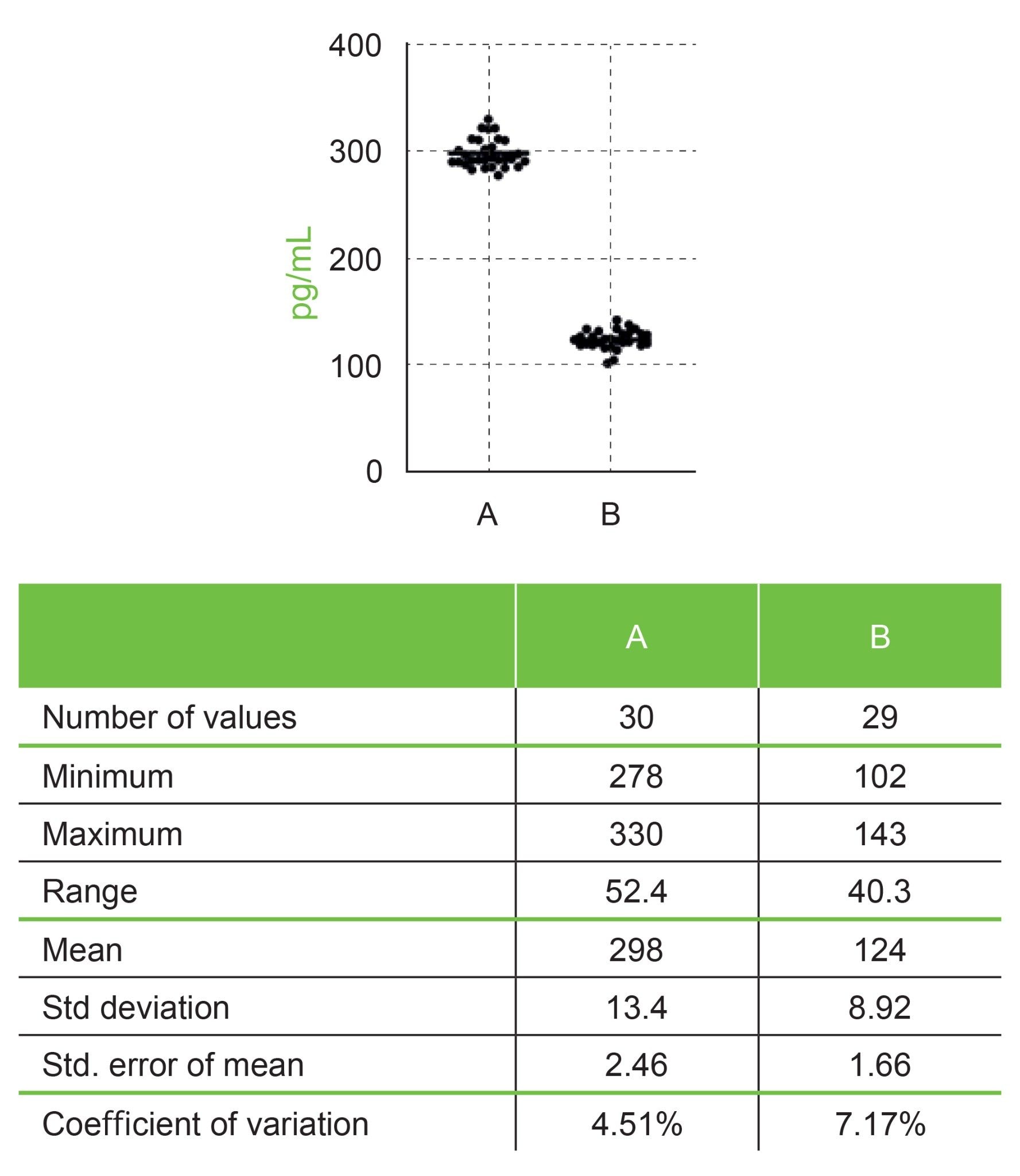 Figure 4. Serum based sample results for Fungitell executed by Andrew+.
The scatter plot contains 30(A) or 29(B) points per sample and the line in each set is the mean. Each sample was run on each of three plates ten times. Each plate was a full execution complete with new standard curve creation. These experiments were executed on the same day across three plates using the same lot of reagents. Note sample B had one data point which was 415 pg/mL, determined to be an outlier and was excluded from this analysis (See text above for more).
Once again these results were excellent with variances across the three plates for the samples ranging from ~4 to 7%. It should be noted one result was excluded from the analysis for sample B as the result was >415 pg/mL. After physically inspecting the plate, understanding the design of the experiment for this sample i.e. single sample transfer tip, single well, same bulk sample, and examining the execution logs from OneLab to confirm no anomalies in sample loading pattern; it was concluded that this was a “hot” well. This value is also a statistical outlier on the plate and across the set.
Conclusion
The results for the standard curve slopes (CV 2.1%, n=8) and intercepts (CV 11.5%, n=8) derived from eight plates collected on two different days had low variance and the R-values for the linear fits at 0.997 or better in each case. These results are equal to or better than the range of variances observed for standard curve results derived from the manual execution of this assay under similar experimental design using the same lots of reagents and materials.
Two sets of glucan containing samples were tested in these experiments. In each case these samples were tested in replicates across three plates on the same day using the same lots of reagents and materials. The variances observed for both of these sample sets were excellent ranging from approximately 4 to 11%. Overall these results demonstrate that Andrew+ Robot driven by the OneLab designed protocol can execute the Fungitell assay within the expected requirements outlined in the instructions for use.
There have been a number of publications describing custom automation processes for the execution of LAL based assays for endotoxin5 and glucan.6 While all have to overcome the same issues regarding consistency and sensitivity to contamination, none have the simplicity and accessibility demonstrated using Andrew+.
References
- Odabasi, Z., Paetznick, V., Rodriguez, J., Chen, E., McGinnis, M., and Ostrosky-Zeichner, L. 2006.Differences in beta-glucan levels of culture supernatants of a variety of fungi. Medical Mycology 44: 267–272.
- De Pauw, B., Walsh, T.J., Donnelly, J.P. et al. 2008. Revised definitions of invasive fungal disease from the European Organization for Research and Treatment of Cancer /Invasive Fungal Infections Cooperative Group and the National Institutes of Allergy and Infectious disease Mycosis Study Group (EORTC/MSG) Concensus Group. Clin. Inf. Dis. 46: 1813–1821.
- Tanaka, S., Aketagawa, J., Takahashi, S., Tsumuraya, Y., and Hashimoto, Y. 1991. Activation of a Limulus coagulation factor G by (1→3)-ß-D-Glucans. Carbohydrate Res. 218:167–174.
- Fungitell Instructions For Use. https://www.fungitell.com/fungitell_assay#Kit_ifu.
- Tsuji KI, Martin PA, Bussey DM. 1984 Automation of chromogenic substrate Limulus amebocyte lysate assay method for endotoxin by robotic system. Applied and environmental microbiology. Sep 1;48(3):550-5.
- Prüller, F., Wagner, J., Raggam, R.B., Hoenigl, M., Kessler, H.H., Truschnig-Wilders, M. and Krause, R., 2014. Automation of serum (1→3)-beta-D-glucan testing allows reliable and rapid discrimination of patients with and without candidemia. Medical mycology, 52(5): 455–461.
Featured Products
720008040, August 2023