For research use only. Not for use in diagnostic procedures.
This is an Application Brief and does not contain a detailed Experimental section.
This application brief demonstrates the desorption of electrospray ionization (DESI) mass spectrometry imaging of lung allograft samples following ex vivo lung perfusion (EVLP) in clinical research, using the Xevo G2-XS QTof Mass Spectrometer.
DESI imaging analysis of donated lung specimen provides highly accurate and robust molecular data with minimum sample handling.
Over the past decade, Mass Spectrometry Imaging (MSI) has been increasingly used by researchers to investigate the distribution of metabolites, drugs, peptides, and proteins in tissue surfaces. The potential for the application of MSI to unambiguously map hundreds of biomolecules in a single analysis has led to this approach being used in research studies of cancer. Recently, there has been a significant increase in the application of Desorption Electrospray Ionization (DESI) as this soft ionization technique can be performed under ambient environmental conditions. Furthermore, it requires little to no sample preparation and is minimally invasive which makes it suitable for direct tissue analysis. DESI-MSI has a potential to provide information about biochemical distribution of molecules after just one measurement in a non-subjective fashion. Therefore, this technique allows researchers to accomplish robust tissue recognition and identification of tissue-specific lipid ion patterns which could, in future, be useful in cancer diagnosis and prognosis at a histology-level.
DESI-MSI is compatible with both the Waters SYNAPT G2-Si and the Xevo G2-XS Mass Spectrometer.
Preceding DESI analysis, donor lung samples are stored at -80 °C. Prior to imaging, samples are removed from the freezer and 10 µm-thick tissue sections are cryosectioned, then thaw-mounted onto glass slides (See Figure 1 for workflow).
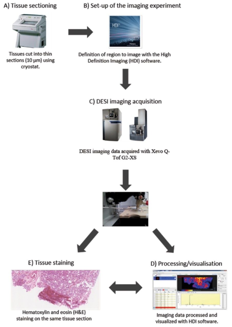
The tissue section is then loaded on the 2D-linear moving stage of the DESI source mounted onto the mass spectrometer. In this case, the Xevo G2-XS Mass Spectrometer was used. This method is also compatible with DESI source on a SYNAPT G2-Si. The area to be analyzed is defined with Waters High Definition Imaging (HDI) Software, version 1.4.
Spatial resolution is adjusted to 100 µm by pre-setting pixel sizes in the x and y directions. Mass spectra are acquired at a 1 scan/second scan rate.
A non-destructive solvent system of 95:5 methanol:water was used at a flow rate of 1.5 µL/min. Nitrogen gas pressure was set to 7 bar.
DESI imaging raw data files are subsequently processed using HDI Software. After DESI imaging, the slides are subjected to traditional H&E staining and scanned using the NanoZoomer S210 scanner. The resulting optical images are coupled with the corresponding molecular images to compare the histopathologic data obtained with the formerly acquired ion images.
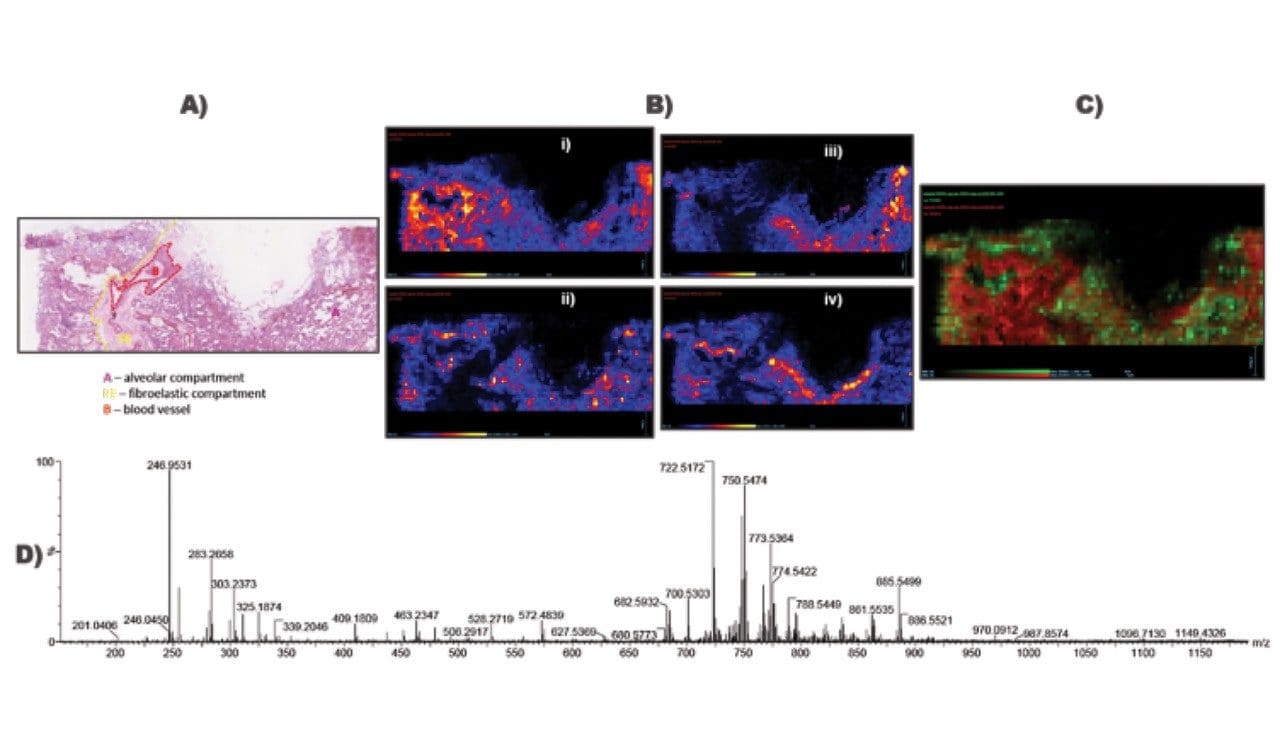
DESI imaging analysis of human lung tissue sections, conducted in both positive and negative ionization modes with a spatial resolution of 100 µm, resulted in a selection of ion images highlighting different tissue compartments, as also observed on the corresponding H&E-stained histological scans and shown in Figures 2 and 3. Overlays of ion images showcasing different molecular distributions were also obtained (Figures 2 and 3).
Utilization of the desorption electrospray ionization technique for mass spectrometry imaging of donor lung specimens carries numerous advantages in Clinical Research:
720005797, June 2017