Drug Metabolite identification relies on confident detection and structural characterization of metabolites. Obtaining high quality data in the presence of diverse and complex matrices (tissue/fluid type/species) can be challenging. It is essential to track and identify these metabolites, especially given the existence of biological mechanisms by which two or more isobaric metabolites can be formed. By measuring CCS accurately it is possible to track these metabolites and glean insight into how the biotransformation has changed the molecular shape. Increasingly, scientists are invoking advanced analytical tools such as ion mobility to help solve these challenges.
Historically, ion mobility-equipped systems required complex and separate drift time enabled software packages for processing and data interpretation. UNIFI Software and the Vion IMS QTof Mass Spectrometer now implement this in a straightforward and user friendly manner, enabling easy access to cleaner data and accurate CCS measurements.
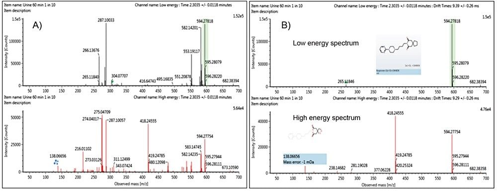
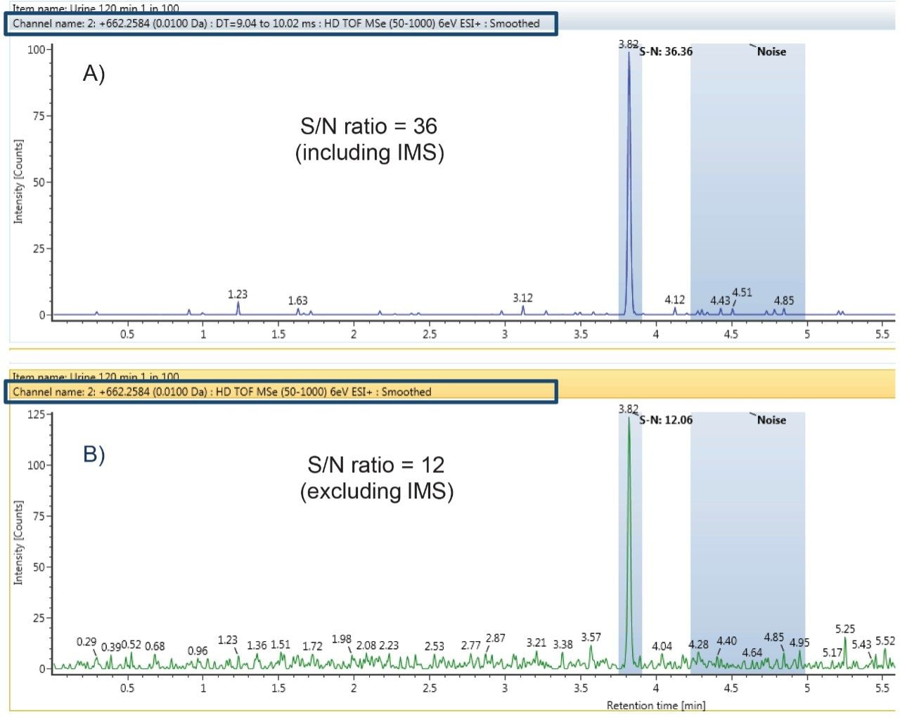
The benefits of ion mobility include: generation of the highest quality data possible with cleaner spectra; access to CCS values for all ions; and the ability to go beyond mass resolution, in order to obtain additional information about metabolite isomers.
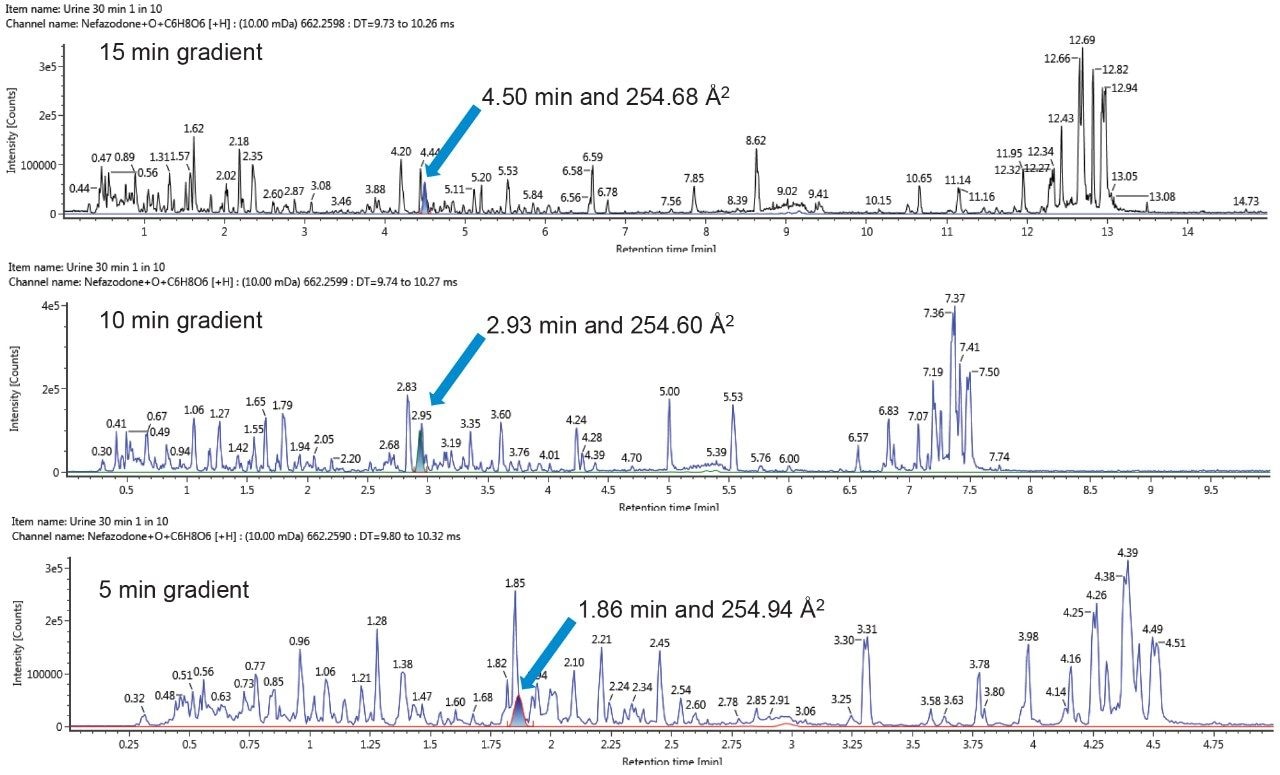
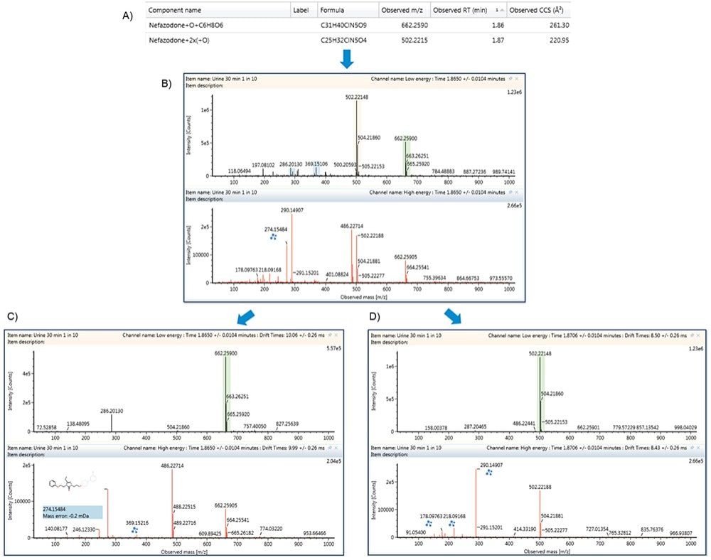
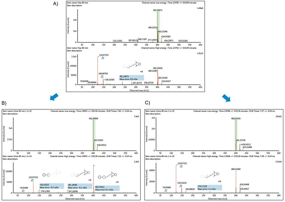
This application note outlines and focuses on the benefits and the ease of use of ion mobility for the application of metabolite identification. Here we highlight a data independent ion mobility data collection approach (HDMSE), to track and resolve, including tracking metabolites across different chromatographic conditions. We include examples of chromatographic co-elution which would otherwise have resulted in mixed product ion spectra.