Rapid High Sensitivity LC-MS/MS Bioanalytical Method for the Simultaneous Quantification of Gefitinib Based PROTACs – 3 and Gefitinib in Rat Plasma to Support Discovery DMPK Studies
본 응용 개요서는 구체적인 실험 내용을 포함하지 않습니다.
Abstract
PROTACs molecules represent a new approach to candidate drug design that can overcome the drug resistance experienced by many small molecule therapies and increase access to previously undruggable proteins, whilst reducing the manufacturing costs, scale-up issues, shelf-life, stability, and storage issues associated with protein biotherapeutics. The accurate quantification of these PROTACs molecules in blood derived fluids is critical to support discovery and development DMPK packages. A rapid (4-minute) LC-MS/MS bioanalytical assay for the quantification of PROTACs-3-gefitinib and gefitinib in rat plasma was developed. The limit of detection (LOD) was determined to be 20 pg/mL for gefitinib PROTACs-3 from 10 µL sample, with a linear dynamic range of 20 pg/mL–1,000 ng/mL. The assay was subjected to a 3-day validation with CV of 5% at the LOD.
Benefits
PROTACs quantification in plasma, LC-MS/MS, DMPK, Bioanalysis.
Introduction
Proteolysis Targeting Chimeras (PROTACs) are a new class of drug molecules which work by mobilizing the ubiquitin–proteasome system to achieve proteasome-mediated degradation of the target protein via the cell machinery. PROTACs consist of three main components i) target binding moiety, ii) linker, and iii) ubiquitin E3 ligase binding moiety, Figure 1. These heterobifunctional molecules function via the binding of the target moiety to the protein of interest (POI) while E3 ubiquitin ligase is simultaneously bound by the other end of the PROTACs molecule. Binding of the POI and the ligase causes ubiquitination of the POI which is then degraded by the cellular ubiquitin-proteosome system, during which the PROTACs molecule is regenerated. These PROTACs can be considered “large small molecules” and as such share many of the attributes of small molecules, such as synthesis scale up, cost of manufacture, shelf life, stability, and route of administration. PROTACs eliminate all functions of the target protein via degradation, hence providing differentiated pharmacology, and do not require target binding moieties that inhibit protein function. They also significantly increase the number of “druggable” proteins, opening up the possibility for new safer medicines.1,2
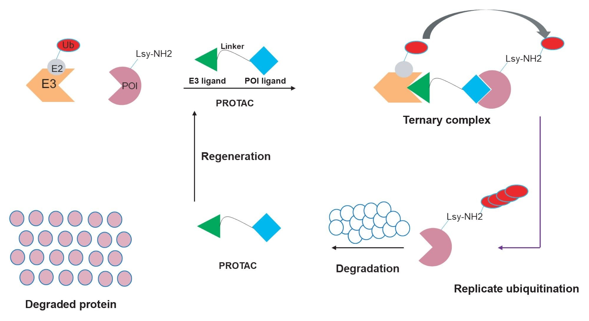 Figure 1. PROTACs catalysed protein degradation.
Gefitinib is a tyrosine kinase inhibitor for the treatment of non-small cell lung cancer, it is a potent epidermal growth factor receptor (EGFR) inhibitor which operates by interrupting cell signalling.3 The pharmacokinetics and in vivo metabolism of gefitinib in the rat and mouse have been previously reported and showed that the compound was well absorbed with peak concentrations occurring at 1 hour, with an estimated oral bioavailability of 50–60%, and a half-life of 3.8 hours (2.6 hours for the IV route).3,4 Previous publications have demonstrated that PROTACs molecules have a long half-life (11–15 hours) with measurable concentrations 48 hours post dose. To facilitate the determination of the pharmacokinetics of gefitinib based PROTACs-3 a high sensitivity bioanalytical method was developed for the simultaneous determination of gefitinib and gefitinib based PROTACs-3 in rat plasma over the concentration range of 20 pg/mL to 1000 ng/mL.
Experimental
Sample Description
Calibration lines were prepared by the dilution of authentic standard of gefitinib and gefitinib based PROTACs-3, Figure 2, in control Wistar rat plasma over the range of 10 pg/mL to 1,000 ng/mL. The methodology was developed and validated in accordance with the FDA Bioanalytical Method Validation Guidance for Industry, May 2018.5 As such the volume of organic solvent from the authentic standard spiking solution was kept below 5% (v/v). Quality control samples (QCs) were prepared in a similar manner from a separate weighing of authentic standards. The plasma samples were prepared by mixing 20 µL plasma with 60 µL acetonitrile containing gefitinib-d6 as the internal standard at a concentration of 50 ng/mL in a 1.5 mL microcentrifuge tubes. The solution was vortex mixed and stored at -20 °C for 1 hour, the sample was then vortex mixed again before centrifugation at 25,000 g for 5 minutes at 4 °C. The resulting sample was transferred to an autosampler vial for analysis. Back calculated standard and QC concentrations were estimated by internal standard quantification using linear regression with a 1/x weighing.
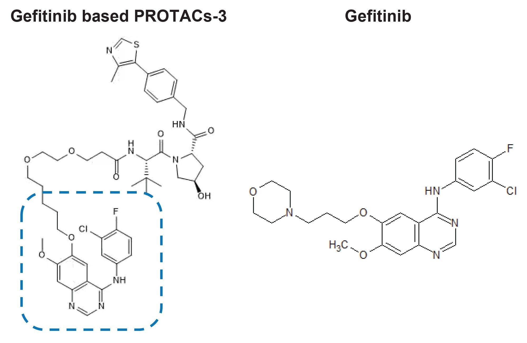 Figure 2. PROTACs-3 Gefitinib (Gefitinib component in dotted line section) and Gefitinib.
Method Conditions
Plasma extracts (2 µL) were analysed using an ACQUITY™ UPLC™ I-Class System connected to a Xevo™ TQ-XS tandem quadrupole mass spectrometer (Waters Corp., Wilmslow, UK). See Tables below for chromatographic, mass spectrometry condition, and informatics employed in this study.
LC Conditions
|
LC system: |
ACQUITY UPLC I-Class |
|
Vials: |
TruView LCMS Certified Clear Glass Screw Neck Total Recovery Vial p/n: 186005669CV |
|
Column(s): |
ACQUITY HSS T3™ C18 2.1 x 50 mm 1.7 µm Column p/n: 186009467 |
|
Column temperature: |
60 °C |
|
Sample temperature: |
10 °C |
|
Injection volume: |
2 µL |
|
Flow rate: |
600 µL/min |
|
Mobile phase A: |
0.1% FA (v/v) in 1 mM aqueous ammonium formate |
|
Mobile phase B: |
0.1% FA (v/v) in ACN containing 1 mM aqueous ammonium formate |
|
Gradient: |
See below |
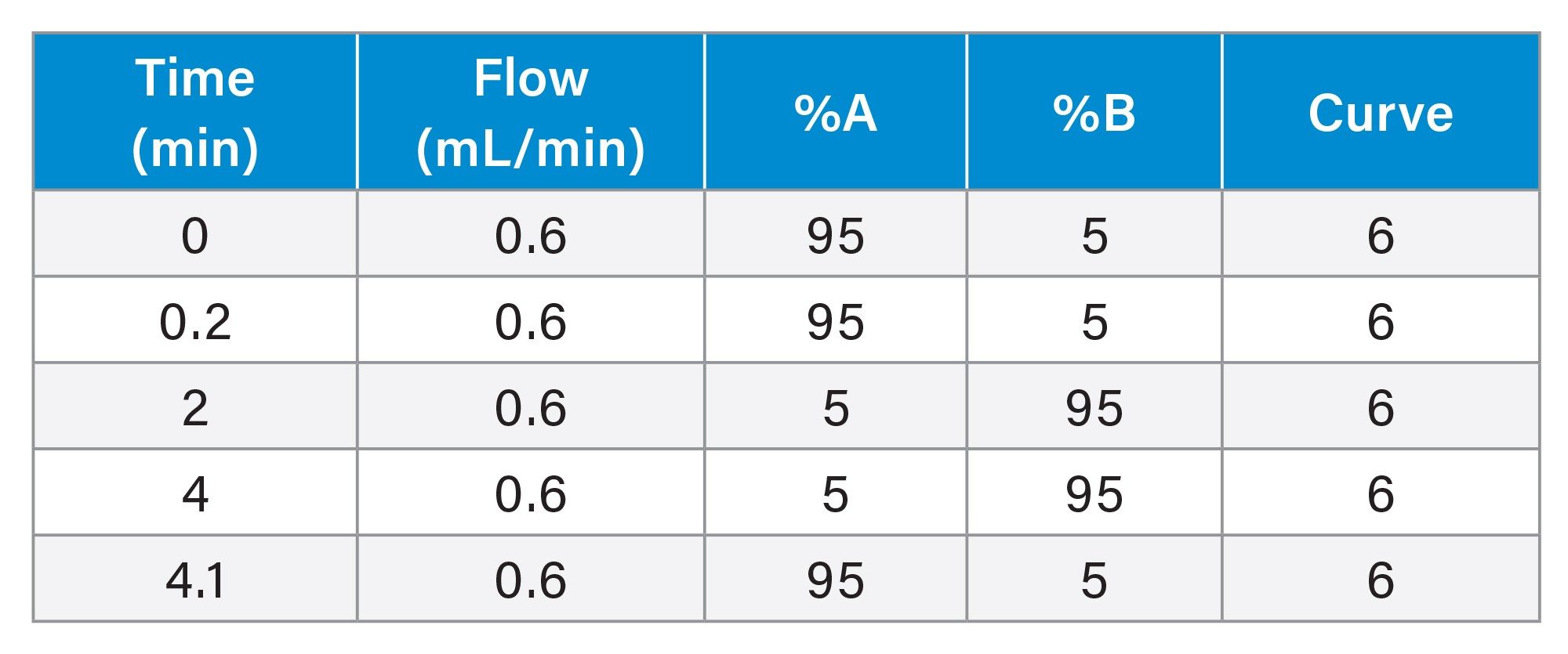
Gradient Table
MS Conditions
|
MS system: |
Xevo TQ-XS Triple Quadrupole Mass Spectrometry |
|
Ionization mode: |
positive electrospray ionisation (ESI+) |
|
MRM transitions: |
|
|
Gefitinib based PROTACs-3: |
m/z=934.33 > 617.34 (CV=60 V, CE=34 eV) |
|
Gefitinib: |
m/z=447.25 > 128.20 (CV=36 V, CE=30 eV) |
|
Gefitinib-d6: |
m/z=453.16 > 134.2 (CV=50 V, CE=48 eV) |
|
Capillary voltage: |
2 kV |
Data Management
|
Acquisition software: |
MassLynx™ V 4.2 |
|
Processing software: |
TargetLynx™ XS |
Results and Discussion
The bioanalytical method was developed for the simultaneous quantification of Gefitinib (C22H24ClFN4O3) and Gefitinib based PROTACs-3 (C47H57ClFN7O8S). The chromatographic separation was optimised to allow for the detection of the dosed compounds and the metabolites of gefitinib and gefitinib based PROTACs-3. The calibration range was evaluated from 10 pg/mL to 1,000 ng/mL in rat plasma.
Mass Spectrometry
The mass spectrometry MRM method parameters for each analyte were evaluated using the MassLynx Intellistart™ software. A 100 nL/mL solution of each analyte (50:50 acetonitrile : water, 0.1% formic acid) was infused, separately, at a flow rate of 10 µL/min (using the on board fluidics of the mass spectrometer). The precursor, productions, cone voltage, and collision energy were evaluated in both ESI+ and ESI- modes. All three analytes gave the strongest response in ESI+ mode, an example of IntelliStart optimisation report for the gefitinib based PROTACs-3 analyte is given below in Figure 3. The results show that the analyte gave rise to a protonated precursor ion with a mass to charge ratio of m/z=934.33, and major fragment ions were detected at m/z=617.34 and 182.06. The cone voltage profile for both analytes was fairly flat optimizing at 88 V for the m/z=617.34 ion, the optimal collision energies were determined to be 34 and 58 eV respectively. The transition m/z=934.17–617.34 was selected for quantification of gefitinib based PROTACs-3 due to its more intense response, gefitinib was monitored using the transitions m/z=447.25–128.20 (CV=36 V and CE=30 eV) and gefitinib-d6 was monitored using the transitions m/z=453.29 to 134.25 (CV=50 V and CE=48 eV). The mass spectrometry acquisition conditions for gefitinib previously reported by Molly et-al., using tandem quadrupole mass spectrometer were employed in the acquisition of the gefitinib concentrations and are listed above in the methodology section.4
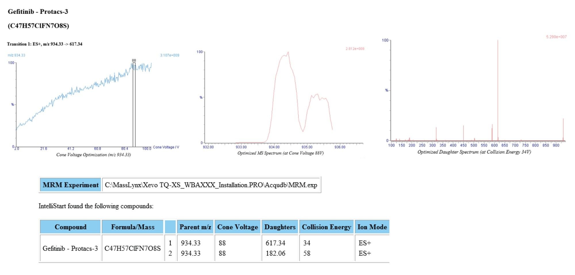 Figure 3. Intellistart Positive ion ESI MRM optimization of Gefitinib based PROTACs-3.
Chromatography
Previous investigation into the in vivo metabolism of gefitinib identified several polar metabolites which required the use of a reversed – phase gradient starting at a low organic composition to prevent co-elution with gefitinib.3,4 Gefitinib based PROTACs-3, however required a high organic concentration in the mobile phase to effect elution. Thus, the chromatography was optimized for the separation of gefitinib, gefitinib based PROTACs-3 and any potential biotransformation’s. The final conditions employed a ACQUITY UPLC HSS T3 Column operated at temperature 60 °C with a gradient of 5–95% acetonitrile – aqueous gradient over 2 minutes with a 2 minute hold at 95% acetonitrile, before returning to the initial conditions at 4.1 minutes, using a flow rate of 600 µL/min. Under these conditions gefitinib and gefitinib based PROTACs-3 eluted with retention times of tR=1.40 and 1.86 min respectively. Figure 4.
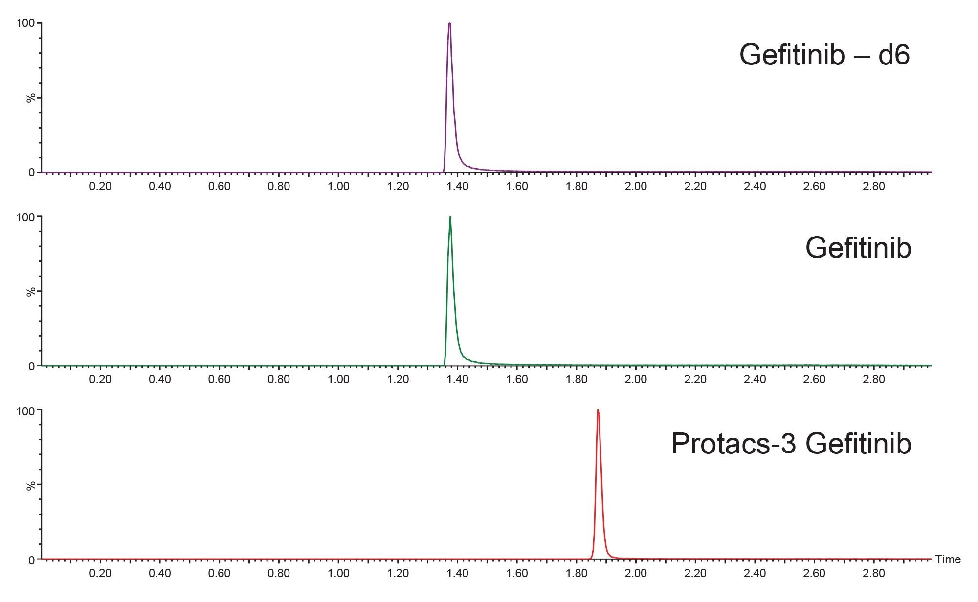 Figure 4. Positive ion LC-MS/MS analysis of gefitinib based PROTACs-3, gefitinib-d6 and gefitinib using a 2.1 x 50 mm ACQUITY HSS T3 1.7 µm C18 Column, maintained at 60 °C and eluted with a 5–95% aqueous – acetonitrile 1 mM ammonium formate, 0.1% formic acid gradient over 2 minutes with a 2 minute hold.
Sample Preparation
The plasma samples were prepared by protein precipitation using 1:3 ratio of sample (20 µL) to organic solvent, as detailed above.
Calibration Range
The lower limit of detection (LLoD) and usable linear dynamic range of the gefitinib based PROTACs-3 method was evaluated over the range of 10 pg/mL to 1,000 ng/mL. The limit of detection in rat plasma was determined to be 20 pg/mL (2.5 fg on column), a representative chromatogram is given in Figure 5. The calibration line was determined to be linear over approximately 5 orders of magnitude (20 pg/mL–1,000 ng/mL) using internal standard quantification and 1/x weighting. A representative calibration line is presented in Figure 6, the correlation coefficient was determined to be r2=0.998, with a negative intercept of -152.165.
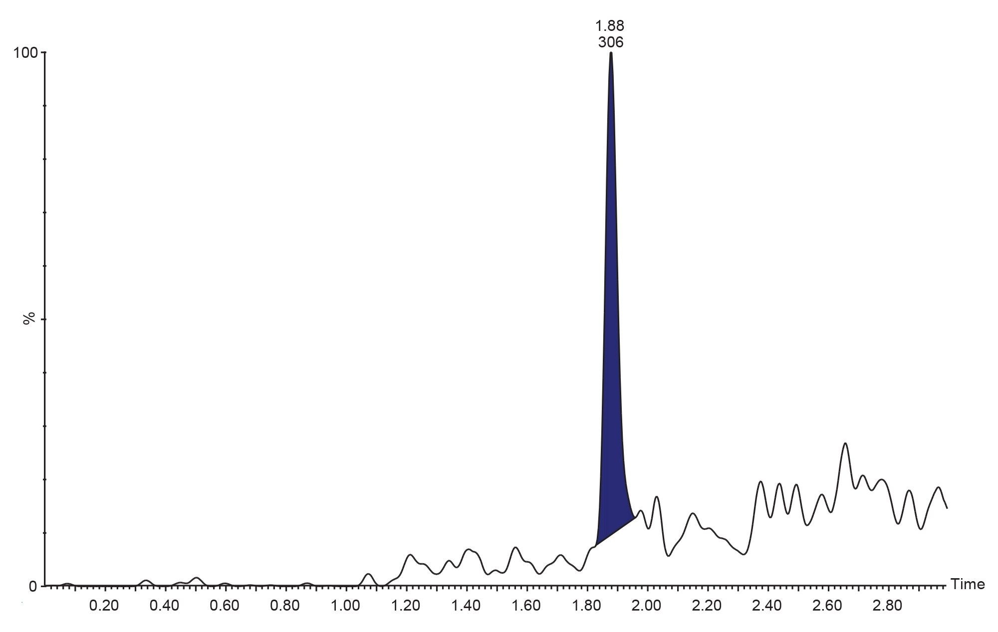 Figure 5. Positive ion LC-MS/MS analysis of gefitinib based PROTACs-3 20 pg/mL standard using a 2.1 x 50 mm ACQUITY HSS T3 1.7 µm C18 Column, maintained at 60 °C and eluted with a 5–95% aqueous – acetonitrile 1 mM ammonium formate, 0.1% formic acid gradient over 2 minutes with a 2 minute hold.
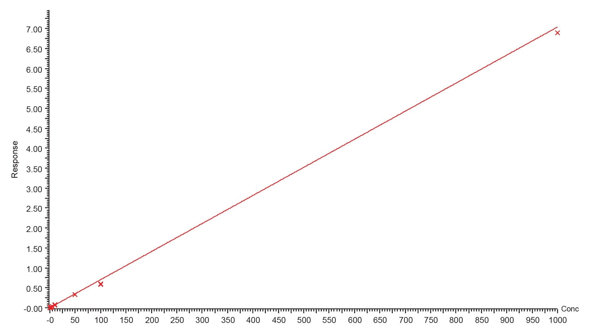 Figure 6. Gefitinib based PROTACs–3 calibration line from 20 pg/mL to 1000 ng/mL.
The limit of detection for gefitinib was determined to be 0.05 ng/mL, with a correlation coefficient of 0.997, a typical calibration line and LOQ standard for gefitinib in rat plasma extract are given in Figure 7.
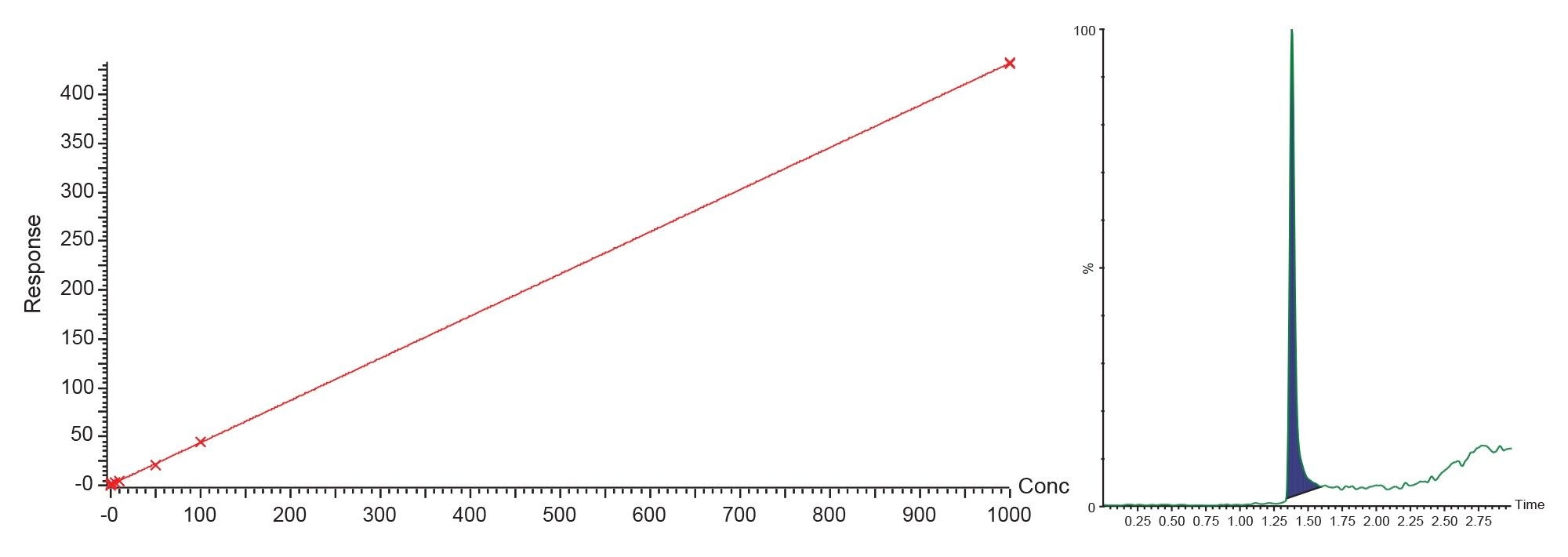 Figure 7. Gefitinib calibration line from 0.5 to 1000 ng/mL and LOQ standard (0.5 ng/mL).
3-Day Validation
Previous studies have shown that gefitinib is well absorbed following PO administration (mouse and rat) with peak plasma concentration being observed at the 1-h time point, with maximum concentration values of 7 µg/mL were obtained for 50 mg/kg dose to the mouse.4 Plasma concentrations rapidly decline with a half-life 3.8-h with virtually no drug being detectable after 24-h post dose. Thus, for a typical pre-clinical safety assessment study where dose levels typically vary from 1–10 mg/kg an analytical concentration range of 0.5–1000 ng/mL was selected for validation, the bioanalytical method for both gefitinib and gefitinib based PROTACs-3 was subject to a 3 day validation. Samples were analysed in three batches over three days in the following order; solvent blank; blank; calibration curve (0, 0.5, 1, 2, 5, 10, 20, 50, 100, 500, and 1000 ng/mL); matrix blank; QCs (3, 75, 800 ng/mL) samples; blank; extracted plasma samples; QCs (3, 75, 800 ng/mL); blank; calibration curve (0, 0.5, 1, 2, 5, 10, 20, 50, 100, 500, and 1000 ng/mL); blank. The three-day validation showed excellent reproducibility and accuracy for both compounds, Tables 1 and 2.
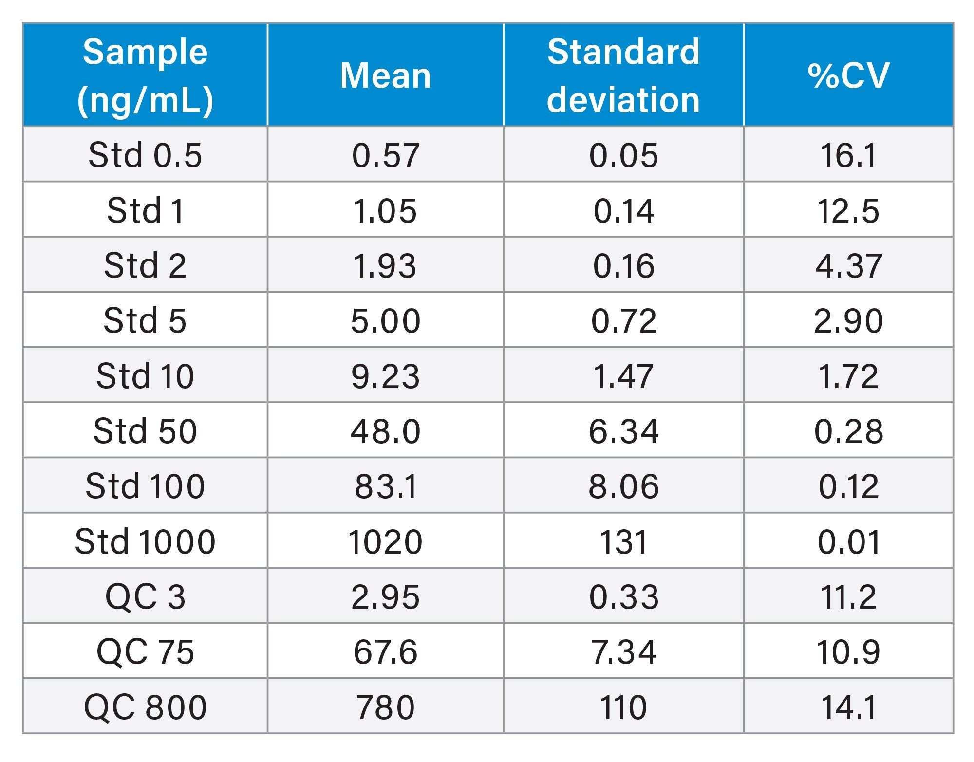 Table 1. Summary of gefitinib based PROTACs-3 quantification in 3-day validation (in total 6 analysis of each standard and QC).
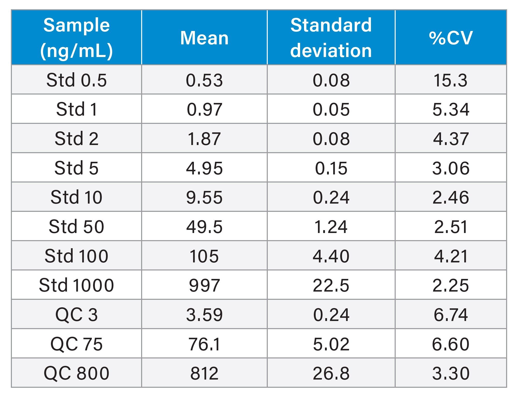 Table 2. Summary of gefitinib quantification in 3-day validation for all data (in total six analysis of each standard and QC).
The freeze-thaw stability of both gefitinib and gefitinib based PROTACs-3 was determined by the analysis of QC samples 3, 75, and 800 ng/mL, the samples were analysed directly following preparation and then following three freeze-thaw cycles at -20°C. The results showed that the measured concentrations % accuracy ranged from 102.9–108.1 for gefitinib to 69.7–109.1% for gefitinib based PROTACs-3.
Conclusion
Proteolysis Targeting Chimeras (PROTACs) use protein targeting ligand linked to a ubiquitin E3 ligase binding moiety to effect proteasome mediated degradation of a target protein and have molecular weights in the 700–1000 Da range. As part of drug discovery and development, the DMPK characteristics of process PROTACs molecules needs to be determined, in a similar manner to all small molecules and biologics; thus a high sensitivity bioanalytical assay is needed. A reversed – phase gradient LC-MS/MS bioanalytical method was developed and validated for the quantification of gefitinib based PROTACs-3 in rat plasma, using the Waters tandem quadrupole mass spectrometer. The limit of detection was determined to be 20 pg/mL (2.5 fg on column) with a dynamic range on 0.02–1000 ng/mL. The method was subject to a three day validation over the range of 1–1,000 ng/mL.
References
- Bhole RP, Kute PR, Chikhale RV, Bonde CG, Pant A, Gurav SS. PROTACs: A Comprehensive Review of Protein Degradation Strategies in Disease Therapy. Bioorg Chem. 2023;139:106720. doi: 10.1016/j.bioorg.2023.106720.
- Békés M, Langley DR, Crews CM. PROTAC Targeted Protein Degraders: The Past Is Prologue. Nat Rev Drug Discov. 2022;21(3):181–200. doi: 10.1038/s41573-021-00371-6.
- McKillop D, Hutchison M, Partridge EA, Bushby N, Cooper CM, Clarkson-Jones JA, Herron W, Swaisland HC. Metabolic Disposition of Gefitinib, an Epidermal Growth Factor Receptor Tyrosine Kinase Inhibitor, in Rat, Dog and Man. Xenobiotica. 2004;34(10):917–34. doi: 10.1080/00498250400009171.
- Molloy B, Mullin L, King A, Gethings LA, Plumb RS, Wilson ID. The Pharmacometabodynamics of Gefitinib After Intravenous Administration to Mice: A Preliminary UPLC-IM-MS Study. Metabolites. 2021 11;11(6):379. doi: 10.3390/metabo11060379.
- https://www.fda.gov/files/drugs/published/Bioanalytical-Method-Validation-Guidance-for-Industry.pdf
720008229, January 2024