• Nota de aplicación

Characterization of Biotherapeutics: ACQUITY UPLC H-Class Bio with 2D Part 3 of 3: On-line Enrichment of Low Abundance Species

Characterization of Biotherapeutics: ACQUITY UPLC H-Class Bio with 2D Part 3 of 3: On-line Enrichment of Low Abundance Species

  • Robert E. Birdsall
  • Vera B. Ivleva
  • Sean M. McCarthy
  • Weibin Chen
  • Waters Corporation

Abstract

The objective of this application note is to demonstrate that the ACQUITY UPLC H-Class Bio System with 2D Technology is well suited for fractionation and enrichment of low abundant species in biological samples. A therapeutic monoclonal antibody, infliximab, was used to demonstrate this.

This comprehensive three-part series demonstrates that the ACQUITY H-Class Bio System with 2D Technology is well suited for the automated fractionation, desalting, and enrichment of biologically complex samples. The ACQUITY UPLC H-Class Bio System with 2D Technology offers an efficient solution with the flexibility needed for increasing informational content in the characterization of biotherapeutics without compromising productivity.

In this third part of the series, we address how to used 2D UPLC with downstream analyses such as peptide mapping, where peaks of interest may be present in low abundance, and how to apply an enrichment process to ensure digest efficiency and success with the subsequent assay.

Benefits

  • Increased productivity through the automation of the fractionation processes using liquid chromatography
  • Improved efficiency through simultaneous quantitation, desalting, and sample enrichment of a biotherapeutic sample
  • Increased sample recovery by minimizing sample preparation and testing aliquots for protein content

Introduction

 Characterization of biotherapeutic proteins often employ separation techniques that incorporate non-denaturing aqueous buffers such as ion exchange chromatography (IEX).1-3 As a result, the low volatility of the mobile phase, and presence of additives such as salt, prevents direct coupling of the separation to mass spectrometry (MS) to elucidate peak identities.4

Parts 1 and Part 2 of this three-part series demonstrated that the ACQUITY UPLC H-Class Bio System with 2D Technology enables the fractionation and desalting of biotherapeutic samples, while facilitating a direct interface for IEX to MS analysis. However, downstream analyses such as peptide mapping still require peaks of interest to be manually collected for enzymatic treatment.

Furthermore, peaks of interest may be present in low abundance, requiring an enrichment process where several fractions are pooled to ensure adequate protein is available for optimal enzymatic digest efficiency and a subsequent assay.5 Factors such as these can require manned instrument operation and negatively impact productivity in the fast-paced pharmaceutical environment.

The ACQUITY UPLC H-Class Bio System with 2D Technology is Waters’ solution for the on-line enrichment of peaks of interest. The 2nd dimension can be used to enrich low abundance fractions where the 2nd dimension column acts as a trapping/desalting column to retain and enrich the protein variant from multiple first dimension heart-cuts. Furthermore, quantitation of desalted/enriched samples can be calculated directly from the peak area rather than testing an aliquot of the enriched fraction, thus allowing for maximum sample recovery with minimal sample preparation.

The objective of this application note is to demonstrate that the ACQUITY UPLC H-Class Bio System with 2D Technology is well suited for fractionation and enrichment of low abundant species in biological samples. A therapeutic monoclonal antibody, infliximab, was used to demonstrate this.

Experimental

LC conditions (enrichment study)

LC system:

ACQUITY UPLC H-Class Bio with 2D Technology

1st dimension pump:

ACQUITY UPLC Quaternary Solvent Manager, ACQUITY UPLC Column manager

2nd dimension pump:

ACQUITY UPLC Binary Solvent Manager, ACQUITY UPLC Autosampler with FTN

Detectors:

(1st dimension) ACQUITY UPLC TUV (2nd dimension) ACQUITY UPLC PDA

Absorption Wavelength:

280 nm

Vials:

Total recovery vial: 12x32 mm glass, screw neck, cap, nonslit (p/n 600000750cv)

Column:

Protein-Pak Hi Res SP, 7 μm, 4.6 x 100 mm (p/n 186004930) ACQUITY UPLC BEH C4, 300Å, 1.7 μm, 2.1 x 50 mm (p/n 186004495)

Column temp.:

25 °C (IEX); C4: 40 °C

Sample temp.:

4 °C

Injection vol.:

2 μL unless otherwise stated

IEX/RPLC Pump Configuration

Quaternary solvent manager:

Flow rate:

0.500 mL/min

Mobile phase A:

100 mM MES monohydrate

Mobile phase B:

100 mM MES sodium salt

Mobile phase C:

1000 mM NaCl

Mobile phase D:

18 MΩ H2O

Auto•Blend Plus setting:

20 mM MES buffer, pH 6.5, 25–65 mM NaCl in 15 minutes

Binary solvent manager:

Flow rate:

0.250 mL/min for heart-cut, otherwise 0.500 mL/min

Mobile phase A:

18MΩ H2O, 0.1% Formic acid

Mobile phase B:

Acetonitrile, 0.1% Formic acid

Gradient:

5–85% B in 10 minutes

LC conditions (peptide analysis)

LC system:

ACQUITY UPLC H-Class Bio

Vials:

Total recovery vial: 12 x 32 mm glass, screw neck, cap, nonslit (p/n 600000750cv)

Column:

ACQUITY UPLC Peptide BEH C18, 130Å, 1.7 μm, 2.1 x 100 mm (p/n 186003544)

Column temp.:

65 °C

Sample temp.:

4 °C

Mobile phase A:

18 MΩ water

Mobile phase B:

Acetonitrile

Mobile phase C:

Acetonitrile, 1 % formic acid, v/v

Gradient table:

Time(min)

Flow rate(mL/min)

A

B

C

D

0.00

0.200

87.0

3.0

10.0

0.0

60.00

0.200

48.0

42.0

10.0

0.0

61.00

0.200

10.0

80.0

10.0

0.0

64.00

0.200

10.0

80.0

10.0

0.0

65.00

0.200

87.0

3.0

10.0

0.0

70.00

0.200

87.0

3.0

10.0

0.0

MS conditions

Instrument:

Xevo G2-S QTof

Capillary:

3kV

Sample cone:

80V

Source temp:

120 °C

Desolvation temp.:

300 °C

Desolvation gas:

800 L/h

Data management

Enrichment study:

MassLynx Software v4.1 (SCN 8.62)

Peptide mapping:

UNIFI Scientific Information System v1.7

The Waters Protein-Pak Hi Res SP, 7 μm, 4.6 x 100 mm, strong cation exchange column (p/n 186004930) and ACQUITY UPLC Protein BEH C4, 300Å, 1.7 μm, 2.1 x 50 mm Column (p/n 186004495) were conditioned prior to use. Chemical reagents were purchased from Sigma Aldrich and used as received. Carboxypeptidase B (p/n LS005305) was purchased from Worthington Labs and used as received. PNGaseF (p/n P0705S) was purchased from New England Biolabs Inc. and used as received. Modified trypsin was purchased from Promega and used as received. The monoclonal antibody infliximab was received at a concentration of 20 mg/mL. Deglycosylation of infliximab was performed overnight (12 hours) at 37 °C using PNGaseF as per the manufacturer’s protocol. After deglycosylation, the sample was divided into two equal aliquots, with one aliquot being treated with Carboxypeptidase B and incubated at 37 °C for 2 hours as per the manufacturer’s protocol. Trypsin digests of enriched fractions were performed overnight at 37 °C as per the manufacturer’s protocol.

Results and Discussion

ACQUITY UPLC H-Class Bio System with 2D Technology featuring heart-cut technology

As described in parts 1 and 2 of this three-part series, the ACQUITY UPLC H-Class Bio System with 2D Technology featuring the heart-cut process is readily deployed with a two-column configuration as shown in Figure 1. With both valves in position 1 (Figure 1A), the flow from both the quaternary solvent manager and binary solvent manger are independent of each other, allowing for independent gradients to be performed on column 1 and 2.

The heart-cut is performed when the valve positions are temporarily switched to position 2 (Figure 1B), combining the flow paths (Figure 1B blue trace) where eluent from column 1 is redirected to column 2. The ACQUITY UPLC Column Manager supports independent valve control as shown in Figure 1C. With the left valve in position 1 and the right valve in position 2 the flow paths of each column are isolated again, with the 2nd dimension column being eluted to waste. This allows for unbound salts to be washed from the heart-cut fraction, which is trapped at the column head of the 2nd dimension column, using the aqueous phase of the 2nd dimension.

Once desalted, the heart-cut fraction can be readily eluted in a mobile phase amendable to down-stream analyses using the ACQUITY UPLC Binary Solvent Manager. With fraction collection occurring post-optical detection, as shown in Figure 1D, peak area can be used for relative quantification of the eluted fraction.

Figure 1. A plumbing diagram of the Waters ACQUITY UPLC H-Class Bio System with 2D Technology and corresponding valve positions to perform a heart-cut are illustrated in A–D. A) Three charge variants (peak 1-3) are separated in the 1st dimension (IEX) using the ACQUITY UPLC Quaternary Solvent Manager to deliver a salt gradient. B) Synchronized valve switching diverts the flow path to the 2nd dimension where a fraction (heart-cut) of peak 2 is directed to the 2nd dimension column (RPLC). C) Independent valve control allows the left valve to re-engage flow to the 1st dimension column while the right valve remains in position 2 where the heart-cut of peak 2 has unbound salt removed under aqueous conditions from the ACQUITY UPLC Binary Solvent Manager. D) The right valve is returned to position 1 after desalting the heart-cut fraction and a standard reversed phase gradient is used to elute the fraction for collection.

On-line enrichment of low abundance species for peptide analysis

Peptide analysis of species such as the acidic variants in the infliximab sample (Figure 2A) can be challenging to characterize due to their low abundance. As an example, the acidic variant eluting at ~6.6 minutes would require approximately 1.0 minute to collect individually for characterization. A 10-μg injection at a flow rate of 0.500 mL/min would result in a fraction concentration of 0.6 ng/μL with the fractionated sample still requiring a buffer exchange, which could lead to additional protein loss.

Low abundance peaks such as these are often manually collected over several runs where the final samples are pooled to ensure adequate protein is available for multiple enzymatic digests. This process requires manned instrument hours and can negatively impact productivity.

The ACQUITY UPLC H-Class Bio System with 2D Technology offers the ideal solution for fractionation and enrichment of biotherapeutics with high protein recovery. Using the column configuration as before, Auto•Blend Plus was used to deliver a salt gradient from 25 mM to 65 mM NaCl in 20 mM MES buffer at pH 6.5, to separate a 3-μL injection of carboxypeptidase B treated sample of infliximab. The acidic variant (peak A) eluting at ~6.6 minutes as shown in the inset of Figure 2A was arbitrarily selected for fractionation and enrichment. To evaluate fraction enrichment of a biotherapeutic using the ACQUITY UPLC H-Class Bio System with 2D Technology, 1 to 5 heart-cuts from sequential 1st dimension separations were retained on the 2nd dimension column and eluted for quantification. The enriched sample was collected post PDA detector to allow for quantification.

For the 1st dimension, a heart-cut was performed from 6.40 minutes to 6.90 minutes as shown in the inset figure of Figure 2A. The heart-cut fractions were retained on the 2nd dimension column using a mobile phase composition of 95 % water, 0.05 % TFA: 5 % acetonitrile, 0.05 % TFA. Enriched fractions were eluted from the 2nd dimension column using a 10 minute gradient from 5% to 85% acetonitrile with 0.05% TFA with manual collection of the eluent occurring from 16–19.5 minutes (Figure 2B).

Figure 2. On-line enrichment of low-abundance species. A) A 3-μL injection (60 μg) of carboxypeptidase B treated infliximab sample separated on a Waters Protein-Pak Hi Res SP strong cation exchange column using Auto•Blend Plus to deliver a 15-minute salt gradient, from 25 mM to 65 mM NaCl, in 20 mM MES buffer at pH 6.8. A heart-cut was performed from 6.40 minutes to 6.90 minutes as shown in the inset figure. B) Heart-cuts were eluted off the 2nd dimension column using a 10-minute gradient from 5% to 85% organic mobile phase. Overlays of chromatographic traces with enrichment of 1 to 5 heart-cuts are shown with their corresponding recovered mass plotted in the inset graph.

For quantification, a six-point calibration plot was constructed (not shown) using the same CPB treated sample of infliximab with mass loads ranging from 1.3 μg to 7.5 μg. As shown in the inset of Figure 7B, the calculated recovered mass is proportional to the number of heart-cuts used in the enrichment process and demonstrates the fidelity of the instrument to deliver a reproducible method for the enrichment of low abundance species.

To demonstrate the applicability of on-line enrichment using the ACQUITY UPLC H-Class Bio System with 2D Technology, the same enrichment process was applied to the main peak (peak D) as shown in Figure 2A and both enriched fractions were subjected to peptide analysis comparison to elucidate difference, if any, contributing to the charge variant.

Enriched fractions were directly transferred to a vacuum centrifuge and dried at 30 °C followed by suspension in 0.100 M Tris buffer, pH 7.6 at a concentration of 1.25 mg/mL and 1.43 mg/mL for peak A (acidic peak) and peak D (main peak), respectively. Samples were enzymatically digested with modified trypsin from Promega as per the manufacturer’s protocol. An ACQUITY UPLC Peptide BEH C18, 130Å, 1.7 μm, 2.1 x 100 mm Column was used for the analysis. 

Three replicate samples were separated with a 13% to 52% gradient using acetonitrile, 0.1% FA v/v, in 60 minutes. The Biopharmaceutical Platform Solution with UNIFI was used for data analysis of the peptide mapping experiments.

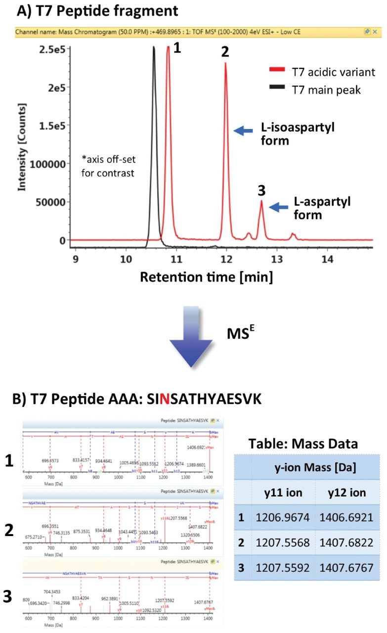
Using a workflow targeted for identification of deamidation events, UNIFI Software was able to identify peptide fragment T7 as shown in Figure 3A as containing a deamidated residue responsible in part for the charge variant of the acidic fraction. The deamidation of asparagine (residue N) undergoes modification from asparagine to a succinimide intermediate that degrades into isoaspartic acid and aspartic acid in a 3:1 ratio.6 These byproducts of the deamidation are observed in the peptide map, following the elution of the unmodified T7 peptide, as +1 Da isoforms.

MSE fragmentation analysis of these species (shown in Figure 3B) revealed that the observed 1 Da mass shifts could indeed be localized to the third residue (N) in the peptide, confirming asparagine deamidation.

This experiment demonstrates that the ACQUITY UPLC H-Class Bio System with 2D Technology is well-suited for fractionation and enrichment of challenging separations encountered during the characterization of biotherapeutics.

Figure 3. Peptide analysis. A) Peptide fragment T7 was identified as containing a deamidated residue (peak 2 and 3) in the acidic variant when compared to the main peak of infliximab. The deamidation of asparagine undergoes modification from asparagine to a succinimide intermediate that degrades into isoaspartic acid and aspartic acid (~ 3:1 ratio).6 B) MSE analysis of the peptides identified a 1 Da shift in the isoapartic and aspartic acid containing deamidated peaks (peak 2 and 3), evident in the y-ions associated with the acidic variant T7 peptide. The characteristic 3:1 ratio of L-isoapartic acid to L-aspartic acid was observed in the BPI profile. A 1 Da shift associated with the deamidation event was observed in y-ions of the acidic fractions amino acid analysis table.

Conclusion

This comprehensive three-part series demonstrates that the ACQUITY H-Class Bio System with 2D Technology is well suited for the automated fractionation, desalting, and enrichment of biologically complex samples. The ACQUITY UPLC H-Class Bio System with 2D Technology offers an efficient solution with the flexibility needed for increasing informational content in the characterization of biotherapeutics without compromising productivity.

References

  1. Liu et al. Chromatographic analysis of the acidic and basic species of recombinant monoclonal antibodies. mAbs. 2012; 4(5): 578–585.
  2. Cheng et al. C-terminal lysine variants in fully human monoclonal antibodies: Investigation of test methods and possible causes. Biotechnology and Bioengineering. 2008; 100(6): 1132–1143.
  3. Brorson, K., Jia A. Therapeutic monoclonal antibodies and consistent ends: terminal heterogeneity, detection, and impact on quality. Current Opinion in Biotechnology. 2014; 30: 140–146.
  4. Annesley, T. Ion Suppression in Mass Spectrometry: Clinical Chemistry. 2003; 49(7): 1041–1044.
  5. Fersht, A. Structure and Mechanism in Protein Science: A Guide to Enzyme Catalysis and Protein Folding, 3rd ed.; W.H. Freeman and Co.: New York, 2000; 103–131.
  6. Geiger, T., Clarke, S. Deamidation, Isomerization, and Racemization at Asparaginyl and Aspartyl Residues in Peptides. The Journal of Biological Chemistry. 1987; 262(2): 785–794.

720005327, March 2015

Volver arriba Volver arriba