• Application Note

Testing for GHB in Hair by GC Tandem Quadrupole MS

Testing for GHB in Hair by GC Tandem Quadrupole MS

  • Marie Bresson
  • Vincent Cirimele
  • Pascal Kintz
  • Marion Villain
  • Timothy Jenkins
  • Jean-Marc Joumier
  • Laboratoire Chemtox
  • Waters Corporation
  • St. Quentin en Yvelines

For forensic toxicology use only.

Abstract

GHB has been misued from its intended therapeutic use by bodybuilders to increase muscle mass; by ravers because it produces euphoria and can balance some side-effects of the drug ecstasy; and by rapists in cases of sexual assault. GHB is detected in blood or urine within 6 to 12 hours of being administered. Hair analysis allows an increase in the detection window depending on the hair length. Recently, hair analysis was used to prove a single exposure. A sensitive, specific, and reproducible method has been developed to detect and quantify GHB in hair by gas chromatography and tandem quadrupole mass spectrometry, GC-MS/MS.

Introduction

Gamma hydroxybutyric acid (GHB) is a substance naturally present within mammal species (Figure 1).

Figure 1. Structure of GHB.

GHB has been used clinically since 1960s as an intravenous anesthetic in the treatment of insomnia, alcohol, and opiate withdrawal syndrome and in cerebrovascular disorders.1

More recently, the molecule has been misued from its intended therapeutic use by bodybuilders to increase muscle mass; by ravers because it produces euphoria and can balance some side-effects of the drug ecstasy; and by rapists in cases of sexual assault.2

GHB is detected in blood or urine within 6 to 12 hours of being administered. Hair analysis allows an increase in the detection window depending on the hair length, with 1 cm corresponding approximately to one month growth. Recently, hair analysis was used to prove a single exposure. For GHB, hair allows analysts to detect the difference between endogenous production and single exposure in sexual assault for example.3

A sensitive, specific, and reproducible method has been developed to detect and quantify GHB in hair by gas chromatography and tandem quadrupole mass spectrometry, GC-MS/MS.

Experimental

Specimens

Hair specimens from forensic cases are used to confirm or disprove a sexual assault under GHB. Specimens are stored at room temperature until analysis.

Extraction

The hair was decontaminated twice using 15 mL of dichloromethane, for 2 min at room temperature.

The segmentation of hair is undertaken to differentiate a single exposure from an endogenous concentration of GHB. Each segment measures 3 mm.

5 to 10 mg of decontamined hair were incubated in 0.5 mL 0.01 NaOH in presence of 10 ng of GHB-d6 used as internal standard overnight at 56 °C. After cooling, the homogenate was neutralized with 60 μL 0.01 HCl, and 2 mL of ethyl acetate were added. After agitation (95 cycles/min for 15 min) and centrifugation, 15 min at 3000 rpm, the organic phase was removed and evaporated to dryness. The residue was derivatized by adding 35 μL BSTFA + 1% TMCS and incubated for 30 min at 70 °C.

GC-MS/MS Procedure

The samples were analyzed on an Agilent 6890 GC coupled to Waters Quattro micro GC tandem quadrupole Mass Spectrometer operated in EI+ mode (Figure 2). Data were acquired and processed with Waters MassLynx Software.

A 1 μL aliquot of extract was injected into the column of a Hewlett-Packard (Palo Alto, CA, U.S.) gas chromatograph (6890 Series). The flow of carrier gas (helium, purity grade 99.9996%) through the column (OV-1 capillary column, 100 % dimethylpolysiloxane, 30 m x 0.25 mm i.d, 0.25 μm film thickness) was 1 mL/min.

The injector temperature was 270 °C. Splitless injection was employed with a split valve off-time of 1.0 min. The column oven temperature was programmed to rise from an initial temperature of 70 °C maintained for 1 min to 160 °C at 15 °C/min, and to 295 °C at 40 °C/min maintained for the final 2 min. The total run time was 12.38 min.

The Quattro micro GC tandem quadrupole Mass Spectrometer was used in electron impact (EI+) mode. The ion source was operated at 180 °C with an electron energy of 70 eV and a filament current of 200 μA. The acquisition mode was multiple reaction monitoring (MRM) at an argon collision gas pressure of 3.00 x 10⁻³ Bar. The electron multiplier was set at 650 V. GHB and GHB-d6, their MRM transitions, collision energies, and retention times are listed in Table 1.

One transition has been chosen to quantify the molecule and the others are used to identify the molecule.

Figure 2. Waters Quattro micro GC-MS/MS System.
Table 1. GHB, GHB-d6 and MRM method parameters. Quantification transition is underlined.

Method Validation

A standard calibration curve was obtained by adding 0.5 ng (0.1 ng/mg), 1 ng (0.2 ng/mg), 2.5 ng (0.5 ng/mg), 5 ng (1 ng/mg), 15 ng (3 ng/mg), 50 ng (10 ng/mg), and 75 ng (15 ng/mg) of GHB in 0.5 mL of NaOH.

Intra-day and between-day precisions for GHB were determined using NaOH spiked with GHB at final concentration of 0.1 ng/mg (n=10).

Relative extraction recovery was determined by comparing the representative peak of GHB extracted at the final concentration of 0.1 ng/mg with the peak area of a methanolic standard at the same concentration (n=3).

The limit of detection (LOD) was evaluated by decreasing concentration of GHB until a response equivalent to three times the background noise was observed for the each transition. For the limit of quantification (LOQ) a response greater than 10 times the background noise is necessary.

Results and Discussion

It is desirable to produce an intense signal that is characteristic for GHB. Selectivity and sensitivity are increased by almost suppressing the noise level. The parent ion of GHB (m/z 233.1 to 233.1 and 131.1) and GHB-d6 (m/z 239.1 to 239.2) correspond to the molecular ion obtained after derivatization. The ion m/z 204.1 (to 162.1) for GHB was chosen based upon criteria of specificity and abundance (Figure 3).

Figure 3. GHB, BSTFA derivatized, EI mass spectrum (70 eV).

The calibration curve corresponds to the linear regression between the peak area ratio of GHB to I.S. and the final concentration of drug for a sample of 5 to 10 mg. Response of GHB was linear in the range 0.1 to 15 ng/mg, and the representative curve for the quantification transition of GHB is illustrated in Figure 4. The correlation coefficient was 0.9997.

Figure 4. Calibration curve for quantification transition of GHB.

The within-batch precision was 9.63% for the quantification transition and the extraction recovery (n=3) was determined to be 49.05%.

The limit of detection was 0.05 ng/mg and the limit of quantification was the first point of the calibration curve, that is, 0.1 ng/mg (Figure 5).

Figure 5. Chromatogram obtained after extraction of GHB at the final concentration at 0.1 ng/mg by the established procedure. Top: GHB-d6 with its daughter ion at m/z 239.2. Middle: quantification transition m/z 233.1 to 233.11. Bottom: two qualifying transitions of GHB.

The analysis of a 2.4 cm strand of hair, obtained from a forensic case, demonstrated the presence of GHB in each segment at the concentration of about 0.17 to 0.23 ng/mg (Figure 6). The chromatogram obtained after the analysis of the last segment is illustrated in Figure 7. This was considered a negative case. A positive case was illustrated in Figure 8.

Figure 6. Hair segmentation from a forensic case.

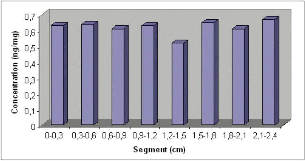
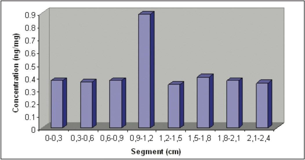
Physiological concentrations of GHB were published by Kintz et al.3 in hair obtained from 24 people. These concentrations were in the range 0.5 to 12 ng/mg. This concentration can be less important, as we have observed with the strand of analyzed hair (0.17 to 0.23 ng/mg).

Everyone has GHB in their organism, and therefore in his or her hair. As a consequence, it is useless to perform assays with the whole strand of hair, considering that the range of concentrations of physiological GHB is relatively important. So, an analysis using segmentation will enable determination due to a single exposure (Figure 8).

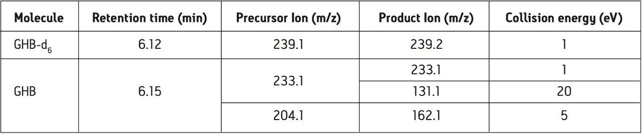
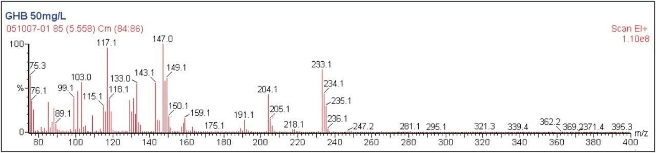
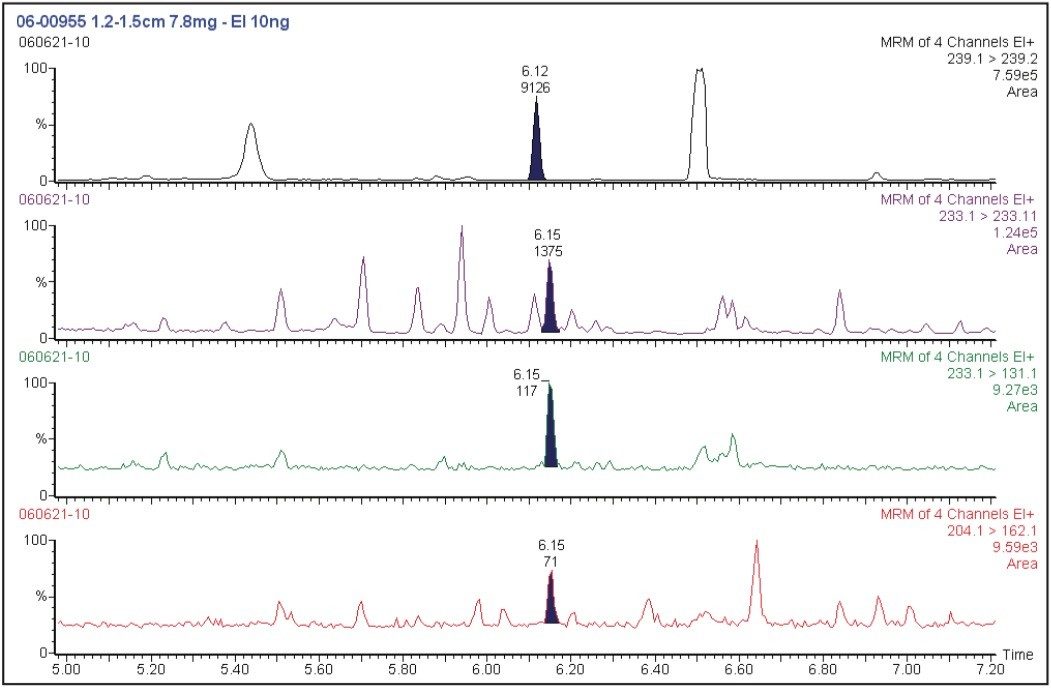
The quantification transition has been chosen according to the peak abundance for GHB. The two other transitions have been chosen based upon criteria of specificity and abundance.

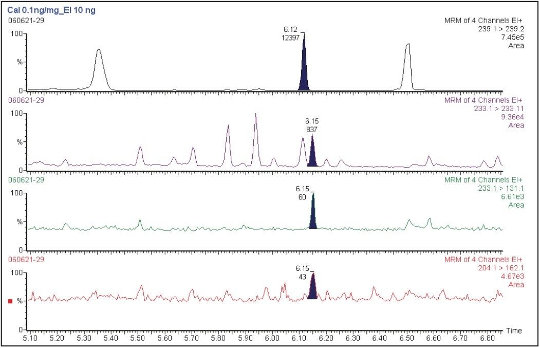
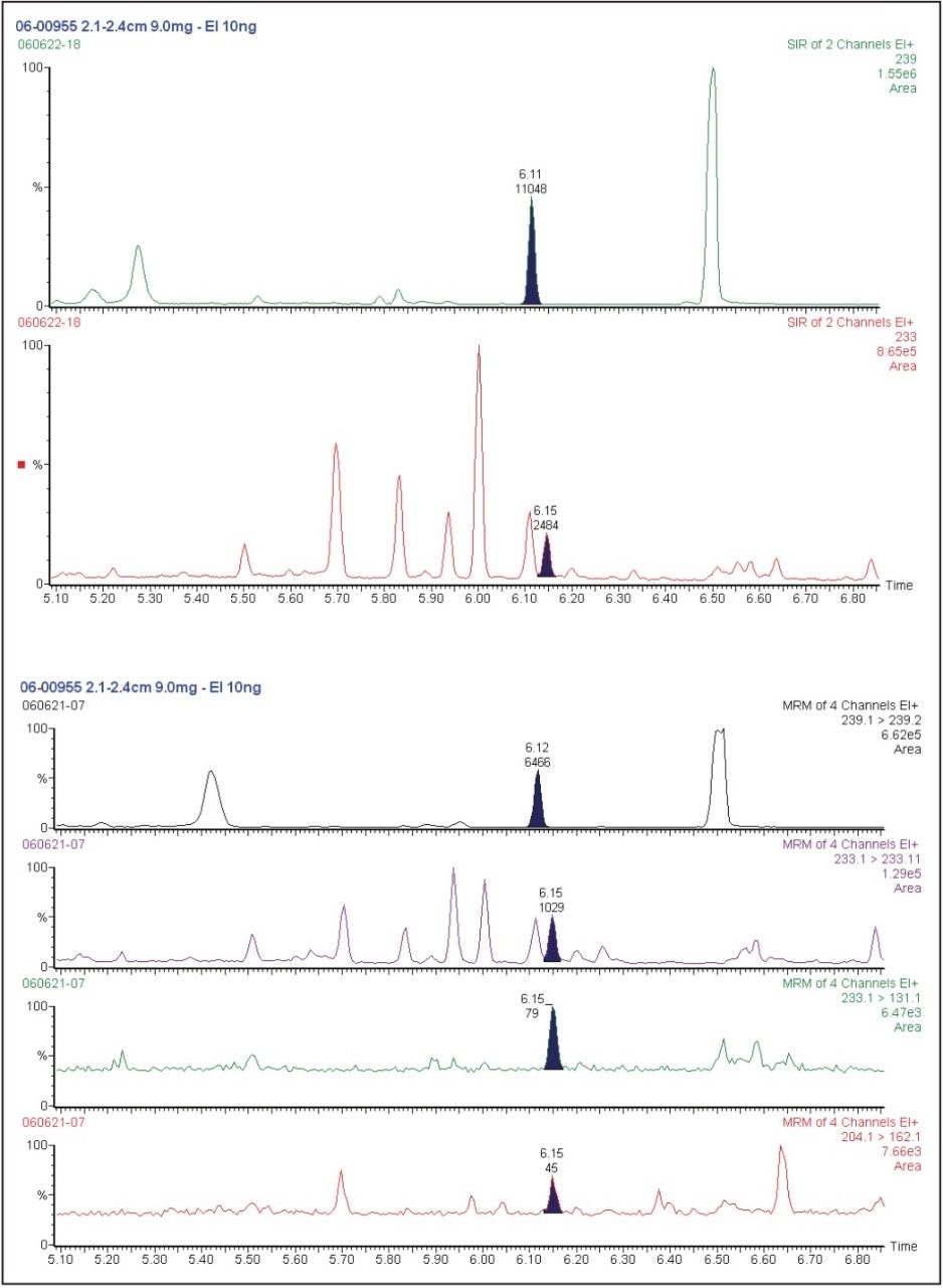
GC-MS/MS appears to be a more useful method for GHB determination in hair than GC-MS. Actually, this method allows us to obtain greater sensitivity and specificity, which is important when trace amounts of analytes are tested. The comparison between MRM and SIR (selected ion recording) modes is shown in Figure 9.

The GC-MS/MS detection in the MRM mode allows us to obtain better chromatograms which are cleaner (lower background noise) and with less interferences than those obtained with GC-MS. This is important as, often, less than 10 mg of hair are analyzed and consequently low concentration of GHB is registered.

Figure 9. Chromatograms obtained in SIR (top) and MRM (bottom) mode of detection after extraction of GHB by established procedure.

Conclusion

The sensitive, specific, and reproducible method appears suitable for the detection and quantification of GHB in human hair.

The segmentation of hair in sexual assault cases is very important to differenciate endogenous production from a single exposure to GHB and to increase the window of detection.

References

  1. 1. P. Kintz, M. Villain, A.L. Pelissier, V. Cirimele and G. Leonetti, J. Anal. Toxicol. 29 (2005) 582.
  2. M.H. Ghysel, Toxicorama XI (1999).
  3. P. Kintz, J. For. Sci. 48 (2003) 195.

Featured Products

720002249, December 2007

Voltar ao início Voltar ao início