For forensic toxicology use only.
A sensitive and reliable method for cannabinoids detection in human whole blood is presented. The procedure involves extraction of a 1 mL specimen acidified with 0.1 mL of 10% acetic acid into hexane/ethyl acetate in presence of deuterated standards. After evaporation to dryness, the drug (Δ9THC) and its metabolites (11-OH-Δ9THC and Δ9THC-COOH) were derivatized by methylation with iodomethane in tetrabutylammonium hydroxyde-dimethyl sulfoxide. The derivates were re-extracted into isooctane before analysis by gas chromatography with tandem quadrupole mass spectrometry (GC-MS/MS). The limit of quantification for each cannabinoid was 0.2 ng/mL, which appears appropriate for use in the investigation of driving under the influence of cannabis, particularly in cases of late sampling.
The detection and quantification of cannabinoids in blood is of major interest in forensic and clinical toxicology.
After smoking, about 18% of Δ9tetrahydrocannabinol (Δ9THC) is absorbed and found in blood. Δ9THC appears rapidly in blood with a peak concentration observed near the end of smoking.1
Oxidative metabolism of Δ9THC leads to formation of the active metabolite, 11-hydroxy- D9tetrahydrocannabinol (11-OH-Δ9THC), that can be further transformed in 11-nor-9-carboxy-Δ9tetrahydrocannabinol (Δ9THC-COOH), as shown in Figure 1.1-2
In suspected cases of driving under the influence of cannabis, to support the observations of the arresting officer, it is necessary to detect, confirm, and quantify the drug and its metabolites in blood.
Most authors, such as Giroud et al.3 or Nadulski et al.,4 have used GC-MS to detect and quantify cannabinoids. In comparison with standard GC-MS procedures, GC-MS/MS will enhance both the sensitivity and the specificity of the analysis. Furthermore, in the case of putrefied blood, GC-MS/MS provides a cleaner chromatogram with less interferences on the peaks of interest.
This paper describes a sensitive, specific, and reproducible method to detect and quantify three cannabinoids in blood.
Specimens were obtained from subjects who died in a car crash where no drug was involved. The specimens were collected in glass vials, and preserved with sodium fluoride or lithium heparinate, that are the mandatory preservatives according to French law. Specimens were stored at 4 °C until analysis. The samples were analyzed on an Agilent 6890 GC coupled to a Waters Quattro micro GC tandem quadrupole mass spectrometer operated in EI+ mode.
The blood sample was extracted according to the procedure described by Kintz and Cirimele5 with some minor modifications (1 mL specimen, dispensable glassware).
Cannabinoids extraction was performed after addition of 50 μL of deuterated internal standards 0.1 mg/L (for a final concentration of 5 ng/mL), 100 μL of acetic acid and 4 mL of hexane/ethyl acetate (9/1, v/v) to 1 mL of whole blood.
After agitation (15 min at 95 cycles/min) and centrifugation (15 min at 3000 rpm), the organic phase was removed and evaporated to dryness.
For derivatization, 200 μL TMAH/DMSO (1/19, v/v) was added to the dried residue. After 2 min at room temperature, 50 μL of iodomethane were added for 15 min at room temperature. The reaction was stopped by adding 200 μL HCl 0.1 M and the cannabinoids were then extracted into 1 mL of isooctane.
After agitation (15 min at 95 cycles/min) and centrifugation (15 min at 3000 rpm), the organic phase was removed and evaporated to dryness. The residue was dissolved in 25 μL of isooctane before injection.
A 1 μL aliquot of extract was injected into the column of a 6890 Series Hewlett Packard (Palo Alto, CA, USA) gas chromatograph. The flow of carrier gas (helium, purity grade 99.9996%) through the column (OV-1 capillary column, 100% dimethylpolysiloxane, 30 m x 0.25 mm i.d., 0.25 μm film thickness) was 1 mL/min.
The injector temperature was 270 °C and splitless injection was employed with a split valve off-time of 1.0 min. The column oven temperature was programmed to rise from an initial temperature of 60 °C maintained for 1 min to 295 °C at 30 °C/min and maintained at 295 °C for the final 5 min. The total run time was 13.83 minutes.
The detector, a Quattro micro GC tandem quadrupole mass spectrometer, was used in electron impact (EI+) mode. The ion source was operated at 180 °C with an electron energy of 70 eV and a filament current of 200 μA. The mode of acquisition was multiple reaction monitoring (MRM) at an argon collision gas pressure of 3.00 x 10⁻³ Bar. The electron multiplier was set at 650V.
Cannabinoids and their MRM transitions, collision energies and retention times are listed in Table 1. For each cannabinoid, one transition has been chosen to quantify the molecule, and the two others are used to identify the molecule.
A standard calibration curve was obtained by adding 0.1 ng (0.1 ng/mL), 0.2 ng (0.2 ng/mL), 0.5 ng (0.5 ng/mL), 1 ng (1 ng/mL), 2 ng (2 ng/mL), 5 ng (5 ng/mL), 10 ng (10 ng/mL), 20 ng (20 ng/mL), and 40 ng (40 ng/mL) of cannabinoid to 1 mL of blood.
Intra-day and between-day precisions for the cannabinoids were determined using negative control blood spiked with cannabinoids at final concentration of 1 ng/mL (n=8).
Relative extraction recovery was determined by comparing the representative peak of cannabinoids extracted from negative control blood spiked at the final concentration of 1 ng/mL with the peak area of a methanolic standard at the same concentration (n=3).
The limit of detection (LOD) was evaluated by decreasing concentration of cannabinoids until a response equivalent to three times the background noise was observed for the three transitions. For the limit of quantification (LOQ) for quantification transition, a response superior to ten times the background noise is necessary.
It is necessary to produce an intense ion signal that is characteristic for the target compound. Using tandem mass spectrometry, selectivity, and sensitivity are increased by almost suppressing the noise level. Under the chromatographic conditions used, there was no interference with the drugs or the internal standards by any extractable endogenous materials present in blood.
Cannabinoids were identified by their retention time (Table 1) and their three specific transitions. The parent ion chosen for the quantification transition of Δ9THC and 11-OH-Δ9THC corresponds to the molecular ion, but not for Δ9THC-COOH; the three transitions for each cannabinoid were chosen based upon criteria of specificity and abundance.
The method is sensitive, specific, and reproducible. A chromatogram obtained from a calibration at 1 ng/mL is shown in Figure 2.
Figure 2. Chromatogram obtained after extraction by the established procedure of 1 mL blood specimen spiked for a final concentration at 1 ng/mL.
Left: Transitions for Δ9THC. Top: Δ9THC-d3; middle: quantification transition; bottom: two qualifying transitions.
Middle: Transitions for 11-OH- Δ9THC. Top: 11-OH- Δ9THC-d3; middle: quantification transition; bottom: two qualifying transitions.
Right: Transitions for Δ9THC-COOH. Top: Δ9THC-COOH-d3; middle: two qualifying transitions; bottom: quantification transition.
The calibration curve corresponds to the linear regression between the peak area ratio of cannabinoids to I.S. and the final concentration of the drug in spiked blood.
The response for the cannabinoids was linear in the range 0.1 to 40 ng/mL. The correlation coefficient was 0.995, 0.998, and 0.996 for Δ9THC, 11-OH-Δ9THC, and Δ9THC-COOH respectively.
The within-batch precisions were 14.1, 11.74, and 10.3% as determined by analyzing eight replicates of 1 mL of blood from the same subject and spiked with a cannabinoids final concentration at 1 ng/mL.
The extraction recovery (n=3) was determined to be 82.4, 74.5, and 93.6% for Δ9THC, 11-OH-Δ9THC, and Δ9THC-COOH respectively. The LOD of the three cannabinoids was 0.1 ng/mL and the LOQ was 0.2 ng/mL. A chromatogram obtained from a calibrator at 0.1 ng/ mL is shown in Figure 3.
Figure 3. Chromatogram obtained after extraction by the established procedure of 1 mL blood specimen spiked for a final concentration at 0.1 ng/mL.
Left: Transitions for Δ9THC. Top: Δ9THC-d3; middle: quantification transition; bottom: two qualifying transitions.
Middle: Transitions for 11-OH- Δ9THC. Top: 11-OH- Δ9THC-d3; middle: quantification transition; bottom: two qualifying transitions.
Right: Transitions for Δ9THC-COOH. Top: Δ9THC-COOH-d3; middle: two qualifying transitions; bottom: quantification transition.
A positive case, in which Δ9THC has been quantified at 0.43 ng/mL, is illustrated in Figure 4.
Figure 4. Chromatogram obtained after extraction by the established procedure of 1 mL of whole blood. Δ9THC was quantified at the concentration of 0.43 ng/mL.
Top: Δ9THC-d3 with its daughter ion at m/z 331.3.
Middle: quantification transition for Δ9THC.
Bottom: two qualifying transitions of
Δ9THC.
In the international literature, there are many publications that describe different methods to extract or to derivatize cannabinoids.
For example, Collins et al.6 used a two-step process of extraction with hexane because Δ9THC and Δ9THC-COOH were in different phases and used BSTFA (N,O-bis(trimethylsilyl)trifluoro-acetamide) for derivation. Giroud et al.3 used a solid phase extraction (SPE) and carried out derivation with iodomethane. Nadulski et al.4 used SPE also, but the derivation step was made with BSTFA.
For the analysis, most authors have used GC-MS. However, Collins et al.6 have used GC-MS/MS but not in the MRM mode.
Moeller et al.7 published a review of the different method of extraction, derivation and analysis used for detection of cannabinoids. In most cases, the LOQ ranged from 0.2 to 3.5 ng/mL.
The method used here for the extraction has been officially appointed as the reference procedure of the French Society of Analytical Toxicology for the determination of impaired drivers in 1996.8
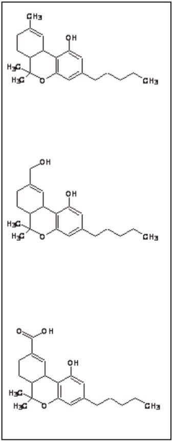
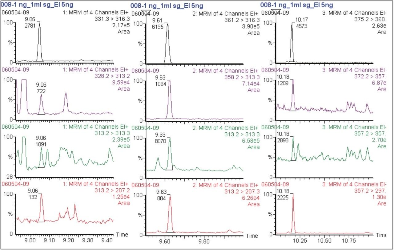
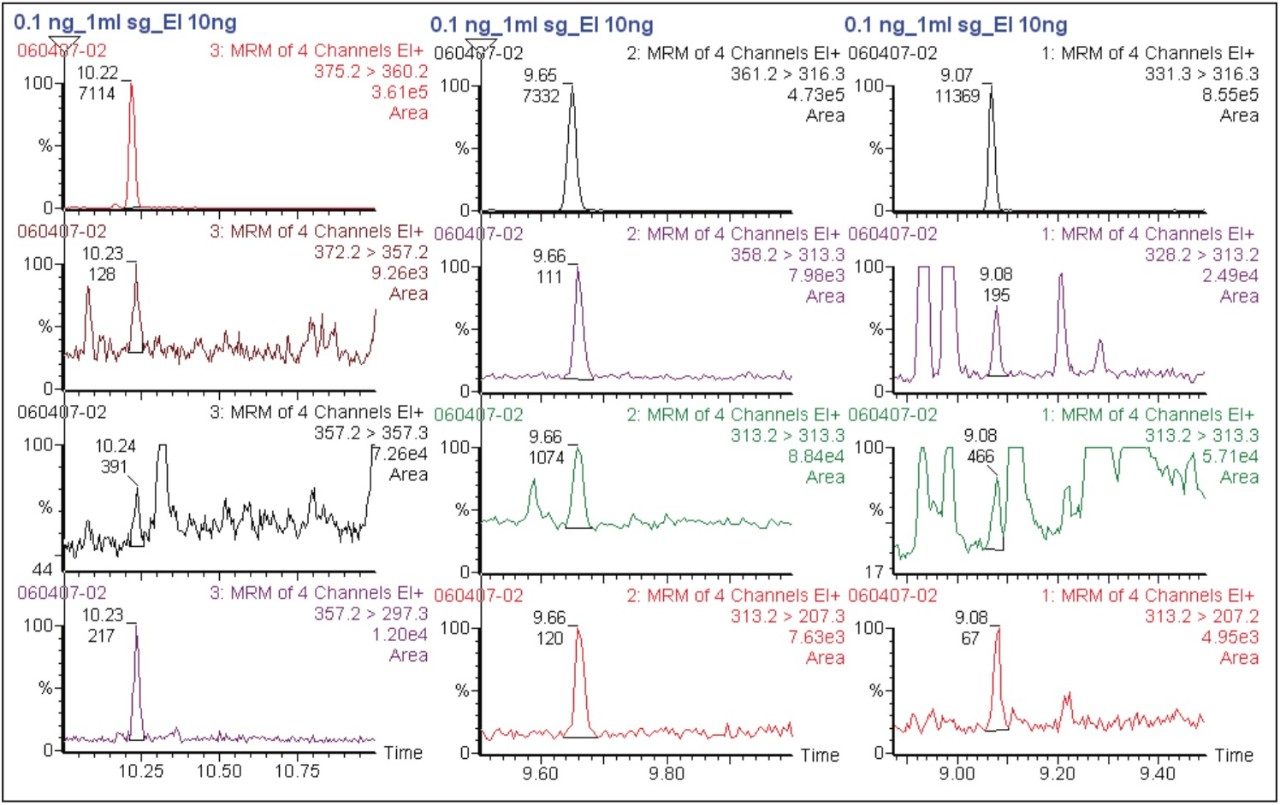
This modified method allows us to obtain higher performances, especially for the LOD that has been reduced at 0.1 ng/mL, instead of 0.4 and 0.2 for Δ9THC and Δ9THC-COOH, respectively. This method allows us to detect and quantify 11-OH-Δ9THC, which was not tested in this previous method.
The interest of GC-MS/MS versus GC-MS is to obtain higher sensitivity and in the possibility of analyzing putrified blood. In this particular situation, cleaner chromatograms were obtained with fewer interferences. The use of disposable glassware was considered as time saving as it was no longer necessary to silanize the vials.
The simultaneous determination of Δ9THC and its metabolites is of importance in both forensic and clinical toxicology to document cannabis impairment. Although GC-MS is the standard procedure, the use of gas chromatography coupled to tandem quadrupole mass spectrometry enhances the analytical security of the test, the LOQ at 0.2 ng/mL appears suitable to extend the time frame of cannabis detection.
720002250, December 2007