• 應用手冊

Best Practices for Oligonucleotide Analysis Using Ion-Pair Reversed-Phase (IP-RP) Liquid Chromatography – Columns and Chemistries 

Best Practices for Oligonucleotide Analysis Using Ion-Pair Reversed-Phase (IP-RP) Liquid Chromatography – Columns and Chemistries 

  • Martin Gilar
  • Nicholas J. Zampa
  • Waters Corporation
需要帮助?
如需详细了解沃特世如何为您抗击新型冠状病毒肺炎(COVID-19)提供助力,请联系

This is an Application Brief and does not contain a detailed Experimental section.

Abstract

Waters presents some critical column and chemistry best practices for performing successful oligonucleotide analysis by ion-pair reversed-phase (IP-RP) liquid chromatography. Ensuring high quality oligonucleotides, either in diagnostic applications or as therapeutic entities, relies on robust analytical methods. For example, in response to the global COVID-19 pandemic, PCR-based diagnostic kits have been developed to detect SARS-CoV-2 genetic code in novel coronavirus patients. Accurate viral detection via PCR requires high quality oligonucleotide primers and probes. Additionally, oligonucleotides are being investigated as therapeutics (including mRNA-based vaccines) for the treatment and prevention of COVID-19.

Benefits

  • Waters BEH-based C18 particles enable high pH and high temperature oligonucleotide separations in LC or LC-MS compatible buffers
  • Eliminate the need for column conditioning, improve analyte recovery, and improve repeatability by minimizing non-specific analyte interactions with metal surfaces with ACQUITY PREMIER Columns with MaxPeak High Performance Surfaces (HPS)
  • Batches are QC tested with MassPREP Oligo Standard to ensure consistency
  • Different particle sizes and column dimensions for preparative or analytical applications
  • Three step method development of <5-minute IP-RP oligonucleotide separations

Introduction

In this application brief, Waters provides a short list of best practices to characterize oligonucleotides by ion-pair reversed-phase (IP-RP) liquid chromatography. These best practices cover column selection, separation temperature, pH optimization, pore size selection, mobile phase choice, rapid method development considerations, purification guidelines, and popular buffer recipes. Following these best practices for oligonucleotide analysis helps ensure robust methods, enabling the delivery of consistently high quality oligonucleotides for therapeutic or diagnostic applications.

The COVID-19 pandemic serves as an example of the importance of oligonucleotides in diagnostic applications. Timely diagnosis of SARS-CoV-2 viral infection remains paramount to successfully managing the novel coronavirus pandemic. As a result, many firms have developed in vitro diagnostic tests to detect the presence of SARS-CoV-2 genetic material. Of the emergency use authorizations (EUA) for in vitro diagnostic tests in the United States, many of them are PCR-based (e.g. qPCR, RT-PCR) tests which detect SARS-CoV-2 viral genetic code.1 Diagnostic kits indicated for the molecular detection of viral genetic code rely on oligonucleotides as primers and probes during PCR amplification and detection. As a result, analytically assessing the quality and ensuring the appropriate purity of oligonucleotide primers and probes is of principle importance for a successful diagnostic test. 

Results and Discussion

Waters created a short list of best practices that have emerged as a result of reviewing method and product development resources for IP-RP oligonucleotide separations. We discuss 7 key practices below.

1)  Consider the use of ACQUITY PREMIER Oligonucleotide BEH C18 Columns with MaxPeak High Performance Surfaces (HPS). PREMIER column hardware built with MaxPeak HPS creates a barrier between the metal surfaces of the fluid path and sample thus minimizing interaction between analytes and column hardware. Employing this technology:

i.  Eliminates the need for time consuming, costly, and transient column conditioning with sacrificial oligonucleotide sample.

ii.  Improves the quality and repeatability of oligonucleotide analyses for both conventional LC-UV applications and, in particular for, sensitive LC-MS analysis. 

For more detailed information, see the Waters Application Note: Improved Chromatographic Analysis of Oligonucleotides with ACQUITY PREMIER Oligonucleotide BEH C18 Columns. 720007012EN.

Figure 1. Comparison of oligonucleotide analyses on conventional versus ACQUITY PREMIER Column hardware. Ten pmol of 15, 20, 25, 30, and 35 mer oligonucleotides (OST MassPREP sample) was injected on new columns (injection #1). Incomplete oligonucleotide recovery is observed on the stainless steel column while nearly complete recovery is obtained with the ACQUITY PREMIER Column. The columns were subsequently conditioned with an injection of a 1 nmol sample of 39 mer oligonucleotide, and the analysis was repeated (chromatograms labeled conditioned). A 25 mM HAA, pH 7 ion pairing mobile phase was used for these experiments.

2)  Perform oligonucleotide separations at elevated pH and temperature for best results.

i.  Elevated temperature (60 °C) prevents the oligonucleotide secondary structure from impacting retention. For CG rich or G-rich oligonucleotides with high degree of secondary structure, it may be necessary to increase column temperature to 80 or 90 °C.

ii.  High pH buffers (pH ≥7) are commonly used in oligonucleotide separations (e.g. TEAA).

iii.  TEA-HFIP was found to be a robust mobile phase offering superior LC-MS sensitivity and resolution across various sized single stranded oligonucleotides.2

iv.  The high pH used for IP-RP oligonucleotide separations renders most common silica-based stationary phases unsuitable.

v.  Waters BEH sorbent technology lends itself well for oligonucleotide separation due to its high pH stability and temperature tolerance.

For more detailed information, see Waters HPLC and UPLC Columns for the Analysis of Oligonucleotides. 720002376EN.

Figure 2. Schematic structure of BEH sorbent. Hydrolytic stability is achieved by bridging ethyl groups. For oligonucleotide analysis, the surface of sorbent is alkylated by C18 functional groups.

3)  Choose a relevant pore size for your oligonucleotide separation. Choosing the correct pore size enables appropriate analyte diffusivity resulting in the best interaction between the oligonucleotide and the ligand. Improved ligand interactions improve peak shape.

 i.  130 Å pore size is ideally suited for single stranded oligonucleotides (2–100 mers).

 ii.  300 Å pore size allows for efficient separation of both single stranded oligonucleotides and longer dsDNA fragments. Even though labeled as a peptide column, scientists have been successful with an ACQUITY PREMIER Peptide BEH C18, 300 Å  Column (186009495, 186009494, 186009493).

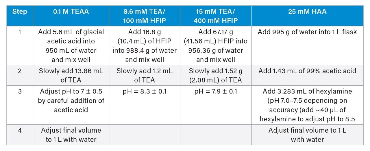
4)  Choose an appropriate mobile phase. See section 7 for buffer recipes.

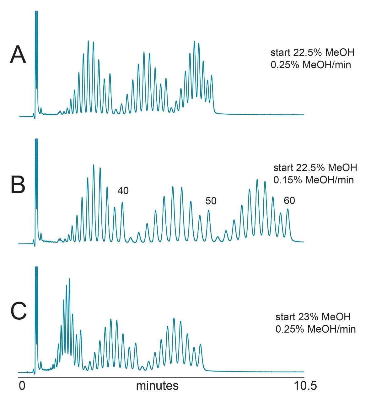
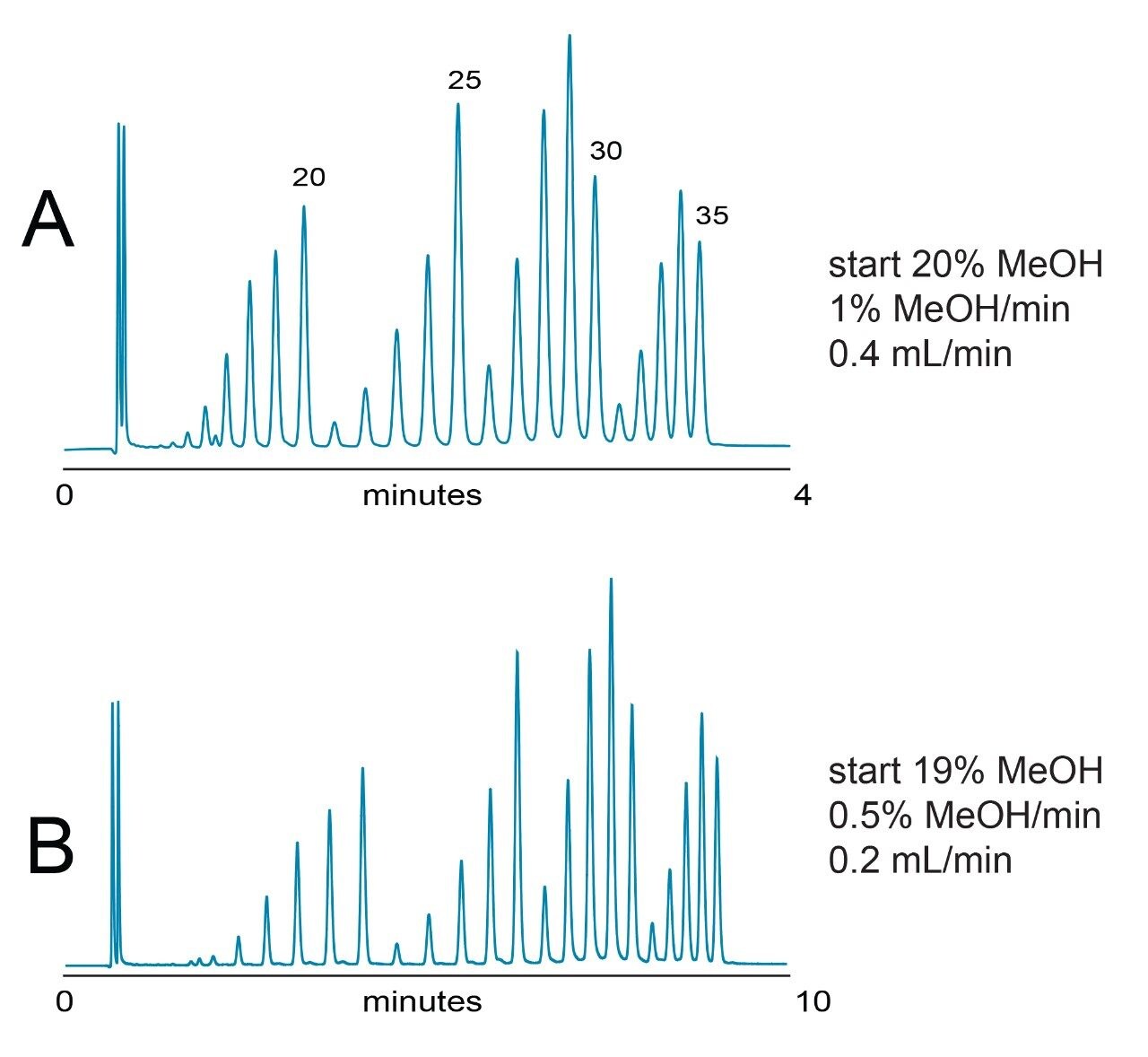
i.  Triethylamine/hexafluoroisopropanol (TEA/HFIP) is MS compatible and has impressive resolving power. Higher TEA/HFIP buffer concentrations improve separation performance. Lower concentrations improve MS sensitivity.

ii.  Hexylammonium acetate (HAA) also offers exceptional resolution and MS compatibility. However, the MS compatibility of HAA is less than that of TEA/HFIP. Use of HAA may result in better separation of labeled oligonucleotides and longer oligonucleotides (>35-mer) compared to TEA/HFIP. This may be relevant if performing only LC analysis.

iii.  Fresh TEA/HFIP and HAA/HFIP mobile phases are critical to good separations. These semi-volatile mobile phases can gradually lose their separation strength and MS spectra become contaminated with alkali ion adducts. For robust day-to-day results, make mobile phases daily or in limited quantities. Upper limit of mobile phase usability is one week.

iv.  Both TEA and HFIP should be prepared in a fume hood, use Waters APC solvent bottle caps to prevent gassing out, and if possible, use a snorkel above the system.

Figure 3. Separation of heteromeric oligonucleotides. The type of ion-pairing mobile phase influences the selectivity of separation. With “weak” IP systems such as TEAA both hydrophobic and ion-pairing interactions participate in separation. With “strong” ion-pairing systems (TEA/HFIP, HAA) the oligonucleotide separation is mostly driven by their charge/length.

For more detailed information, see the below Waters Application Notes

  • UPLC-MS Separation of Oligonucleotides in Less than Five Minutes: Method Development. 720002387EN.
  • Evaluation of Alternative Ion-pairing Reagents in the Analysis of Oligonucleotides with the ACQUITY QDa Detector. 720005830EN.
  • Hexylammonium Acetate as an Ion-Pairing Agent for IP-RP LC Analysis of Oligonucleotides. 720003361EN.

5)   Quick three step method development for <5-minute IP-RP oligonucleotide separations.

i.   Identify suitable initial gradient strength or start with a scouting gradient. With 15 mM TEA/400 mM HFIP ion pairing system, an example scouting gradient may be:

  • Start at 20% MeOH, perform a 1%/min MeOH gradient with 0.2 mL/min flow rate (0.2 mL/min is useful to enhance column efficiency for macromolecules).

ii.   Adjust gradient slope to achieve desired separation (shallower gradients increase resolution, but increase time needed for analysis). Adjust the starting percentage of MeOH to reduce the time of analysis as needed. Extend the MeOH gradient time as needed until the target oligonucleotide and impurities are eluted. High organic flush is then inserted to elute highly retained components (often non-oligonucleotide components) and minimize carryover. Target oligonucleotides should elute during the gradient and not in the high organic flush.

iii.   If speed is important, speed up the separation by increasing the flow rate while proportionally reducing gradient time (constant gradient column volume). Selectivity of separation should not change while minimal loss in resolution can be observed. When driving towards sensitivity or a need for optimal resolution, use lower flow rates.

  • 0.4 mL/min is a good compromise between speed and analytical performance. For high-throughput separations a 0.8 mL/min flow rate is recommended (1–3 min separation time, 2.1 mm column I.D).
  • For more detailed method development considerations for XBridge OST Columns (e.g. starting gradient slopes, buffer formulations, and column considerations) see Waters 715001476.

Figure 4. Separation of 30 to 60 nt oligodeoxythymidines using 2.1 x 50 mm, 1.7 μm ACQUITY UPLC OST C18 Column.

Figure 5. Separation of 15 to 35 ntoligodeoxythymidines.

For more detailed information, see the below Waters Application Notes:

  • UPLC-MS Separation of Oligonucleotides in Less than Five Minutes: Method Development. 720002387EN.
  • UPLC Separation of Oligonucleotides: Method Development. 720002383EN.
  • XBridge OST C18 Method Guidelines. 715001476.

6)   Guidelines for oligonucleotide purification using XBridge Oligonucleotide BEH C18 Columns.

i.   XBridge Oligonucleotide BEH C18, 130 Å Columns are the preferred offering for detritylated oligonucleotide purifications due to the availability of column sizes designed to meet lab-scale isolation requirements.  

ii.   The choice of XBridge Oligonucleotide C18 Column dimension and operating flow rate depends primarily on the scale of the synthesis reaction mixture.

  •  For example, a 4.6 × 50 mm column containing XBridge Oligonucleotide BEH C18, 130 Å, 2.5 μm material is an excellent selection when oligonucleotide mass loads are less than or equal to 0.2 μmol.
  •  Selection of the appropriate column size for oligonucleotide sample load is recommended to maximize component resolution and recovery of the target product from non-desired failure sequences. See Table 1 for mass loading guidelines.
Figure 6. HPLC purification of a synthetic 21-mer oligonucleotide. Sample concentration was 2.8 nmol/μL, with on-column loading ranging from 1.4 to 140 nmol. 

*Values are approximate and vary depending on oligonucleotide length, base composition, and “heart-cutting” fraction collection method used.

Table 1. XBridge OST C18 Column selection guide for detritylated oligonucleotide purification.

7)   How to make select IP-RP buffers (1 liter)

i.   Perform all work in a fume hood.

ii.   Filter all mobile phases through a solvent compatible, 0.45 µm membrane filter and store in bottles that are clean and particulate free.

  • These are all recipes for mobile phase A. Mobile phase B is generally a mixture of organic (e.g. ACN, MeOH) mixed with mobile phase A at a suitable percentage (e.g. 20–50%). The higher concentration buffer (15 mM TEA/400 mM HFIP) can be used for separations involving G-rich oligonucleotides. The lower strength buffer (8.6 mM TEA/100 mM HFIP) is often enough for routine detriylated oligonucleotide LC-MS applications.

Conclusion

In support of customers working on oligonucleotides, Waters offers these critical best practices to ensure consistent high-performance IP-RP liquid chromatographic separations for oligonucleotide analysis. Robust methods are critical to delivering high quality oligonucleotides in diagnostic or therapeutic applications. For example, high quality oligonucleotide primers and probes are essential for accurate PCR-based diagnostic assays, including those for COVID-19.

References

  1. In Vitro Diagnostic EUAs – Test Kit Manufacturers and Commercial Laboratories Table [Internet]. Washington (DC). FDA. [cited 2020 Jun 1]. Available from: https://www.fda.gov/medical-devices/emergency-situations-medical-devices/emergency-use-authorizations#covid19ivd.
  2. Gilar M, Fountain KJ, Budman Y, Holyoke JL, Davoudi H, Gebler JC. Characterization of Therapeutic Oligonucleotides Using Liquid Chromatography with On-line Mass Spectrometry Detection. Oligonucleotides. 2003 13(4):229–243.

720006948, Revised December 2020

回到頁首 回到頁首