• Application Note

Analysis of Deamidation and Oxidation in Monoclonal Antibody using Peptide Mapping with UPLC/MSE

Analysis of Deamidation and Oxidation in Monoclonal Antibody using Peptide Mapping with UPLC-MSE

  • Hongwei Xie
  • Martin Gilar
  • John C. Gebler
  • Waters Corporation

Abstract

This application note demonstrates that UPLC-MSE is a suitable tool for characterizing PTMs in monoclonal antibodies. Using UPLC-MSE with the ACQUITY UPLC and SYNAPT MS systems, the identification of peptide sequences and determination of site-specific moditications can be accomplished in a single LC run because of available peptide fragmentation information provided by MSE.

Introduction

Monoclonal antibodies (mAb) are an important class of protein therapeutics. The mAb production in recombinant expression systems is often a source of post-translational modifications (PTMs), such as various degrees of glycosilation, oxidation, and deamidation.

Asparagine (N) deamidation and methionine (M) oxidation are two common modifications. Deamidation can diminish the activity and stability of an antibody.

Sulfur-containing methionine is a major target for oxidation. Studies1-3 on mAb revealed oxidation during storage. In this application note, we demonstrate the use of data-independent acquisition mass spectrometry, MSE, with UltraPerformance LC (UPLC) for profiling of N-deamidation and M-oxidation sites in mAb after prolonged storage.

Unmodified peptides were resolved from site-specific deamidated isoforms using a Waters ACQUITY UPLC System and identified by MSE. The identities of modified “PENNY” peptides were further confirmed by spiking the sample with synthetic peptide standards. Relative quantitation of the modifications was estimated for both N-deamidation and M-oxidation identified from this antibody.

Experimental

Sample preparation and UPLC-MSE experimental procedure were identical to a previous description.4 Briefly, the antibody was digested with enzyme trypsin after reduction with dithiothereitol and alkylation with iodoacetamide. The resulting peptide mixture was separated using an ACQUITY UPLC System and detected by MSE on a SYNAPT MS System. In order to profile substoichiometric modifications in the antibody, 120 pmol of freshly-prepared tryptic digest was injected for the UPLC-MSE analysis.

The acquired data were processed by IdentityE Software of ProteinLynx Global SERVER 2.3. The processed data4,5 were searched against a database consisting of light and heavy chain sequences of the antibody, with trypsin specificity and one optional miscleavage. Cysteine (C) carbamidome-thylation, asparagine (N) deamidation, glutamine (Q) deamidation, and methionine (M) oxidation were allowed as optional modifications in the search.

UPLC-MSE experiments for synthetic peptides (as listed in Table 1, purchased from Biomatic, Toronto, Canada) were performed using the same experimental conditions as for the digest. The identities of these peptides were confirmed by MSE spectra. Their retention time (RT) is recorded in Table 1.

Table 1. UPLC elution order of synthetic peptides.

Results and Discussion

The features and operational aspects of UPLC-MSE have been described previously.4,5 Briefly, protein digest is separated by UPLC and on-line detected by MSE. Two sets of MS data are collected in parallel: low-energy (MS) and elevated-energy (MSE) chromatograms. The collected data are combined for identification of peptides with the help of sequence database searching analysis. The MSE acquisition is data independent, which ensures sampling of low-abundance peptides and substoichiometric PTMs. The obtained spectra of such peptides allow for identification of peptide modifications. In this study, we focus on characterization of two major degradation pathways: deamidation and oxidation in mAb the antibody.

In order to profile the modifications, the obtained UPLC-MSE data were searched against light and heavy chain sequences of the antibdy with N/Q-deamidation (+0.98 Da) and M-oxidation (+16 Da) as optional modifications. The profiling returns 10 modified peptides, including eight deamidated and two oxidized peptides. The modification type, site, relative concentration, and RT of identified modified peptides are listed in Table 2. For comparison, the corresponding unmodified peptides are also included in the Table. All the MSE spectra of modified peptides were successfully validated.

Table 2. Modification type, site, and relative concentration of modified peptides Identified from the antibody.

Of the two detected M-oxidations (Table 2), about 5% of M255 in the heavy chain was oxidized. The elution pattern and MSE spectra of peptide T21 before and after M255 oxidation is shown in Figure 1. Peptide T21 with M255 oxidation elutes about 4 min earlier than the peptide without modification, because M-oxidation increases the hydrophilicity of peptides.

Figure 1. Elution pattern and MSE spectra of peptide T21 in heavy chain before and after M255-oxidation. Y ions marked in red indicate the M-oxidation.

N-deamidations occur at different levels (Table 2). Each N-deamidation results in two isobaric modified isoforms, isoaspartic acid and aspartic acid, which can’t be differentiated by MS. However, N-deamidated peptide with isoaspartic acid elutes earlier than its counterpart with aspartic acid, as confirmed by spiking the sample with synthetic isoaspartic and aspartic isoforms of deamidated peptide standards (see the elution order and RT in Table 1). The elution order is in agreement with literature data.

The peptide T37 of heavy chain with “PENNY” motif has been well studied in literature and suggested as the peptide most susceptible to deamidation in mAb.6,7 The studies indicate that  deamidation occurs on the first two N sites (N387 and N392). In this study, we show separation of six deamidated products (Figure 2). They are identified (see MSE spectra in Figure 3) to be “PENNY” peptide with isoaspartic acid (isoD) (Peak 2), aspartic acid (D) (Peak 4) and succinimide intermediate (Suc) (Peak 6) of N387 deamidation, as well as newly-found products with deamidation on both N387 and N392 sites (Peak 5), and with deamidation on Q389 (Peak3). N392- deamidated product co-eluted with Q389-deamidated product. This was identified by examining the isotopic patterns of y-series ions in MSE spectrum of peak 3 (data not shown), and further confirmed by RTs when spiking the corresponding synthetic standards in the sample (Table 1).

Figure 2. Elution pattern and MS spectra of “PENNY” peptide T37 in Heavy Chain before and after deamidation. Top – Elution pattern; Bottom – MS spectra.
Figure 3. MSE spectra of Peaks 1, 2 (or 4), 5, and 6 in Figure 2. Y ions marked in red confirm the sequence modifications.

The quantitative results listed in Table 2 show that N387 is the dominant deamidation site of the “PENNY” peptide in this antibody. The total deamidation rate of the “PENNY” peptide was ~ 53.6%.

Comparing the deamidation rate of the “PENNY” peptide with the deamidation of peptides T10 (~ 78.5%) and T6 (~ 51.7%) of heavy chain, we found deamidation on N387, N84, and N55 sites in heavy chain are the major degradation pathways of this antibody.

Conclusion

The results demonstrate that UPLC-MSE is a suitable tool for characterizing PTMs in monoclonal antibodies. MSE ensures sampling of low-abundance components and acquires indiscriminately MSE spectra, enabling accurate identification of modified peptides in an unbiased, reproducible manner. The specific conclusions from this study:

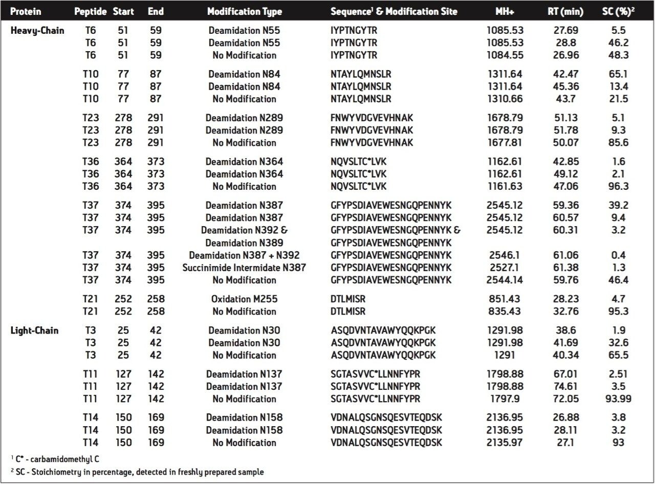
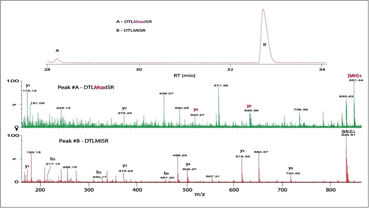
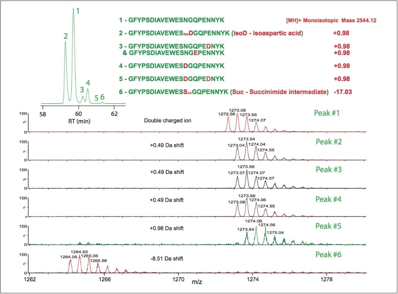
1. UPLC-MSE is capable of separating, identifying, and quantifying modified peptides and isoforms

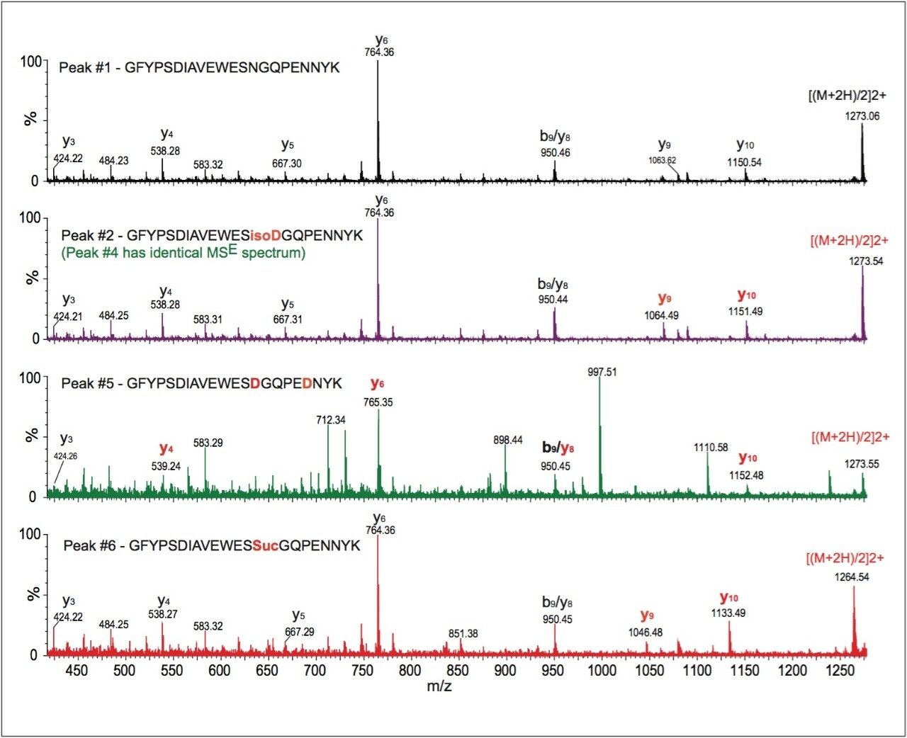
2. The high mass resolution and high mass accuracy of the SYNAPT MS System ensures confident identification of modifications with small mass shift (e.g., N-deamidation with 0.98 Da mass difference) and modified isoforms

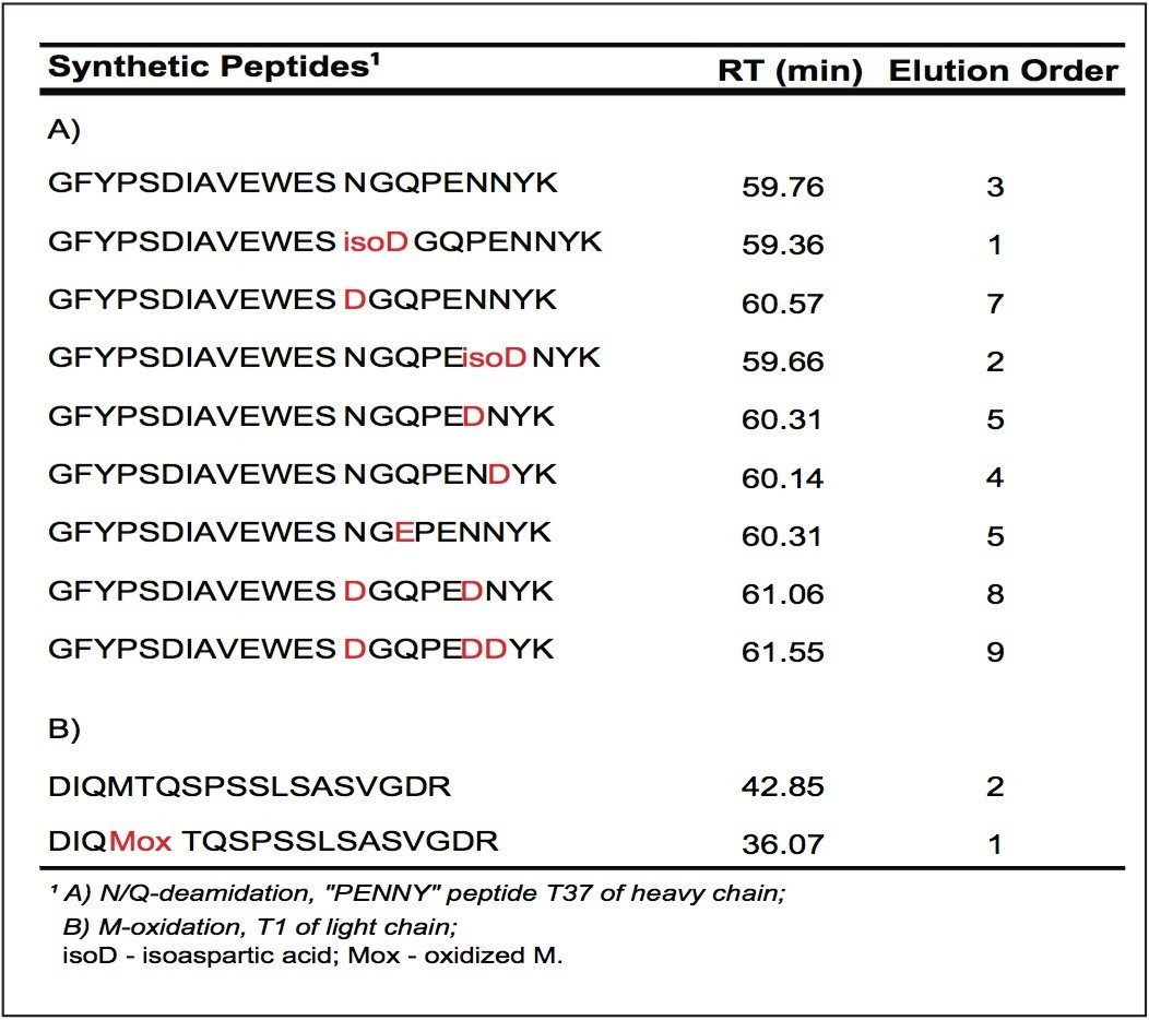
3. Synthetic peptides are helpful for determining modified isoforms and are required for confirmation

In a previous study4, we have demonstrated that UPLC-MSE is able to provide high sequence coverage mapping of mAb tryptic digest, with 97% sequence coverage for both light and heavy chains of the antibody. Therefore, UPLC-MSE and SYNAPT MS system is an advanced platform for characterization of recombinant proteins, such as monoclonal antibodies.

In current LC-UV/MS peptide mapping methods, the identification of peptide sequences and determination of site-specific modifications typically require multiple tandem mass spectrometry experiments (either DDA MS/MS or targeted MS/MS). The methodology reported here achieves both goals in a single LC run because of available peptide fragmentation information provided by MSE.

Peptide mapping with UPLC-MSE improves the analytical efficiency of peptide characterization.

References

  1. Kroon DJ, Baldwin-Ferro A, Lalan P. Identification of sites of degradation in a therapeutic monoclonal antibody by peptide mapping. Pharm. Res. 1992; 9: 1386.
  2. Liu H, Gaza-bulseco G, Xiang T, Chumsae C. Structural effects of deglycosylation and methionine oxidation on monoclonal antibody. Mol. Immunol. 2008; 45: 701.
  3. Liu H, Gaza-bulseco G, Sun J. Characterization of the stability of a fully human monoclonal IgG after prolonged incubation at elevated temperature. J. Chromatogr. B: Anal. Technol. Biomed. Life Sci. 2006; 837: 35.
  4. Xie HW, Gilar M, Gebler JC. High Sequence Coverage Mapping Tryptic Digest of a Monoclonal Antibody with UPLC-MSE. Waters Application Note. 2009; 720002821en.
  5. Xie HW, Gilar M, Gebler JC. Characterization of Protein Impurities by Peptide Mapping with UPLC/MSE. Waters Application Note. 2009; 720002809en).
  6. Chelius D, Rehder DS, Bondarenko PV. Identification and Characterization of Deamidation Sites in the Conserved Regions of Human Immunoglobulin Gamma Antibodies. Anal. Chem. 2005; 77: 6004.
  7. Harris RJ, Shire SJ, Winter C. Commercial Manufacturing Scale Formulation and Analytical Characterization of Therapeutic Recombinant Antibodies. Drug Dev. Res. 2004; 61: 137.

720002897, January 2009

Back To Top Back To Top