In this application note, we demonstrate the utility of UPLC-MSE for identification and quantification of covalent modifications in a yeast alcohol dehydrogenase (ADH) digest.
Protein-based pharmaceuticals are an important class of biotherapeutic compounds. Although proteins are relatively stable molecules, they are subject to a variety of covalent modifications that occur during manufacturing, formulation, and storage. Modifications have the potential to affect activity and safety of protein drugs. Effective monitoring of them is imperative for ensuring the quality of therapeutic proteins.
Liquid chromatography (LC)-based peptide mapping is suitable for detection of covalent modifications. However, for identification of site-specific modifications, tandem mass spectrometry (MS/MS) sequencing is required. This is especially the case when several possible sites in the peptide can be modified.
In data-dependent acquisition (DDA) LC-MS/MS sequencing, multiple experiments usually need to be performed to elucidate the structure of peptides in protein digests. Detailed DDA LC-MS/MS analysis of peptide maps is a time-consuming task.
Recently, we have demonstrated that peptide mapping with UltraPerformance LC (UPLC) and data-independent acquisition mass spectrometry (UPLC-MSE) is effective for characterization of protein maps,1–3 providing high sequence coverage and assignment of nearly all LC peaks in the map. The parallel and unbiased dataindependent acquisition of precursor and fragmentation data by MSE 4–5 overcomes the limitations of DDA and also delivers reliable quantitative data.
In this application note, we demonstrate the utility of UPLC-MSE for identification and quantification of covalent modifications in a yeast alcohol dehydrogenase (ADH) digest. Two common modifications, methionine (M) oxidation and asparagine (N) deamidation, are examined. Modified peptides were clearly distinguished from lowabundant peptides originating from impurity proteins or unexpected peptides resulted from non-specific digestion.
Sample preparation and UPLC-MSE experimental procedure were similar to previous descriptions.1–2 Briefly, ADH was digested with enzyme trypsin after reduction with dithiothereitol and alkylation with iodoacetamide. The resulting peptide mixture was separated using ACQUITY UPLC System and detected using MSE on a SYNAPT MS System. Freshly prepared tryptic digest (120 pmol) was injected for the UPLC-MSE analysis. The analysis was repeated three times.
LC system: |
Waters ACQUITY UPLC System |
Mobile phase A: |
0.1% formic acid in water |
Mobile phase B: |
0.1% formic acid in acetonitrile |
Column: |
ACQUITY UPLC Peptide Separation Technology C18, BEH 300Å, 2.1 x 100 mm, 1.7 μm |
Column temp.: |
40 °C |
Gradient: |
0–50% B in 90 min |
Flow rate: |
0.2 mL/min |
Detection: |
MSE |
The acquired data were processed by IdentityE Software, part of ProteinLynx Global SERVER 2.3. The processed data1–2 were first searched against a yeast database (containing 6139 open reading frames) with trypsin specificity and one potential miscleavage. Then, the data were searched again against the ADH1 sequence with no enzyme specified. N-deamidation, M-oxidation, N-terminal acetylation and C-carbamidomethylation were allowed as variable modifications.
In order to profile sub-stoichiometric modifications in the protein, 120 pmol ADH tryptic digest was injected for the UPLC/MSE experiments. Figure 1 shows an example UPLC-MSE chromatogram.
The collected MSE data were first searched against a yeast database in order to verify the identity of ADH1 and search for potential impurity proteins. Target protein ADH1 and two impurity proteins, ADH5 and Ylr301wp, were found. The concentration of impurity proteins, normalized to ADH1, was 5% and 0.74% for ADH5 and Ylr301wp, respectively.
Subsequently, the MSE data were searched again against a truncated database containing only ADH1 sequence. The search was purposefully relaxed (no enzyme specificity) to allow for identification of non-specifically cleaved and modified peptides. The ADH1 sequence coverage was 98%.
Table 1 shows sequences of eight modified amino acid sites located in seven tryptic peptides, including N-terminal acetylation, three oxidized methionines and four deamidated asparagines. The modification type, site, and stoichiometry as well as retention time (RT) of identified modified peptides are presented. The comparisons of MSE spectra of peptide T12 with and without M-oxidation, and peptide T22 with and without N-deamidation, are shown in Figures 2 and 3, respectively, demonstrating confident identification of the modifications.
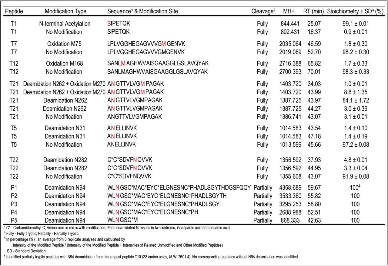
The relative quantification of modified peptides calculated from MS signal intensity (Table 1) shows that over 99% of N-terminal of ADH1 were found to be acetylated. Three methionines in the protein sequence were found to be oxidized to a relatively low degree, less than 2% for M75 and M168 sites, and approximately 10% for M270 site. In addition to M-oxidation, the peptide T21 was nearly completely deamidated at N262 site (97%), present in four isoforms as shown in Table 1. Similarly, high percentage of deamidation was observed for N94 site.
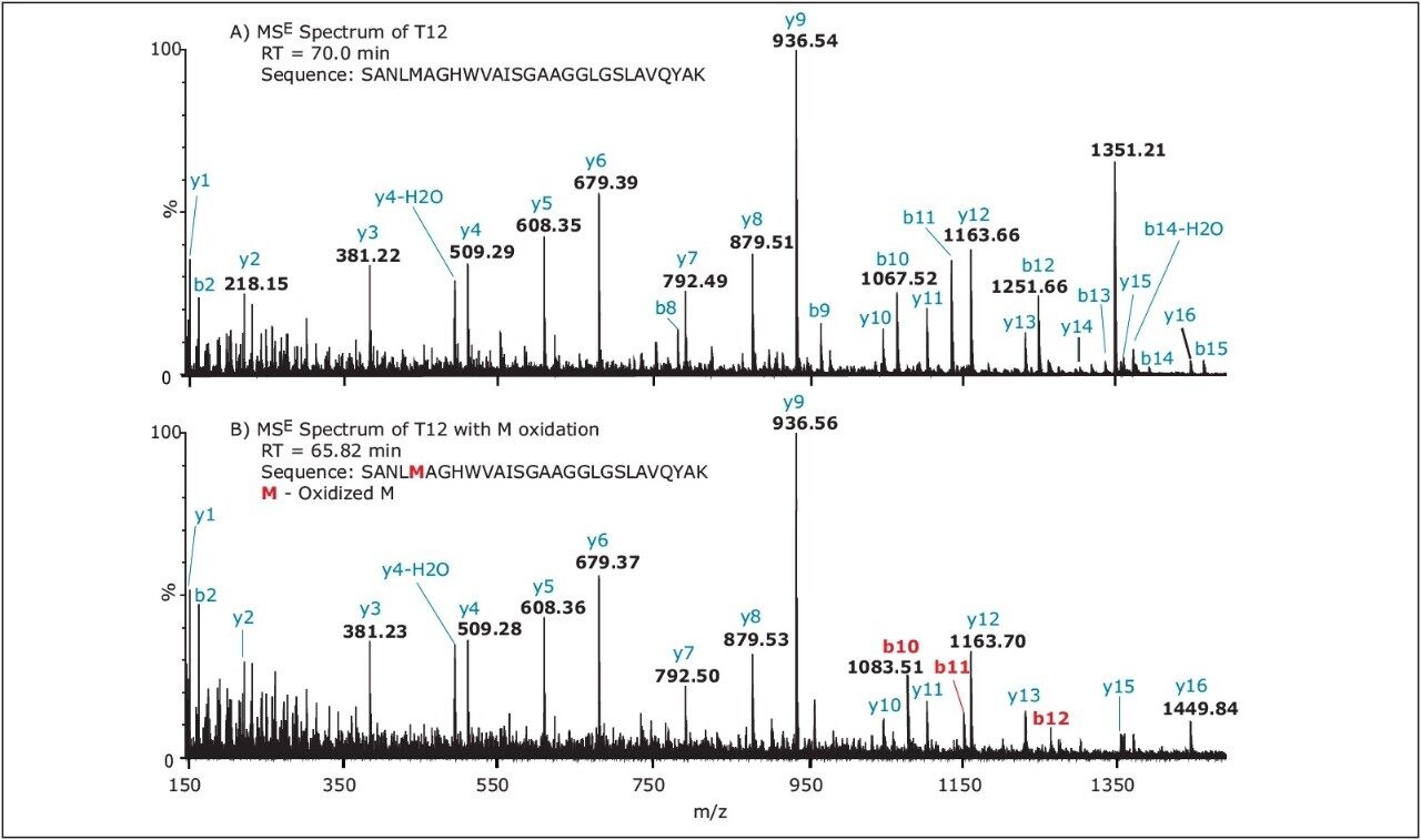
Although the tryptic peptide T10 (consisting of 69 amino acids, molecular weight 7601.4) was not detected, a series of partiallytryptic N94-deamidated peptides covering part of the missing T10 sequence were found (P1, P2, P3, P4, P5 as shown in Table 1). The non-deamidated versions of these peptides were not detected. Finally, the deamidation of N31 and N282 sites was found to be less than 10%. Peptide RT shift upon modification can be used as an additional confirmation of sequence modification. In general, N-terminal acetylation increases, while M-oxidation decreases the peptide retention. The RT of N-terminal peptide T1 shifted from 16.37 min to 25.07 min after N-terminal acetylation. In contrast, the RT shifted from 52.7 min to 46.59 min for peptide T7 and from 70.0 min to 65.82 min for peptide T12 after M-oxidation in these peptides.
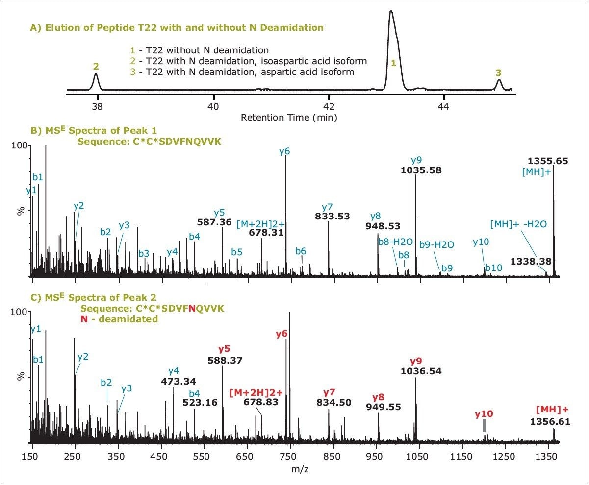
In the case of N-deamidation, the RT trends are more complex because of the presence of two product isoforms: isoaspartic acid and aspartic acid. Usually, N-deamidated peptide with isoaspartic acid elutes earlier and the other deamidated one with aspartic acid later than the unmodified peptide. There are exceptions to this rule, but the retention order observed here was always the isoform with isoaspartic acid < aspartic acid isoform.
The retention pattern of unmodified peptide T22 and its two N282-deamidated isoforms is presented in Figure 3A. Since the two deamidated isoforms are isobaric (both +0.98 Da mass difference from the unmodified T22), they cannot be distinguished from MS or MSE data. In such cases, the UPLC separation and RT information of these peptides are important for the identification. However, MSE can easily differentiate N-deamidated peptide from unmodified peptide, as shown in Figure 3, with a high mass resolution and high mass accuracy platform such as the SYNAPT MS System.
The results presented here demonstrate application of UPLC-MSE for characterization of protein peptide maps. UPLC-MSE was successfully used for identification and quantification of modifications in ADH1. The stoichiometry of modifications ranged from 1% to 99%. In conclusion, peptide mapping with UPLC-MSE allows for:
UPLC-MSE, combining here the ACQUITY UPLC and SYNAPT MS systems, meets requirements for robust and flexible methods that are needed to monitor safety and stability of biopharmaceutical proteins.1-3 This technology is a fitting choice for biopharmaceutical research and development laboratories. Since it is suitable for establishing protein sequence and characterizing protein covalent modifications and impurities within a single UPLC run, the method will expedite the recombinant protein drug development and manufacturing processes, and reduce their cost.
720003009, April 2009