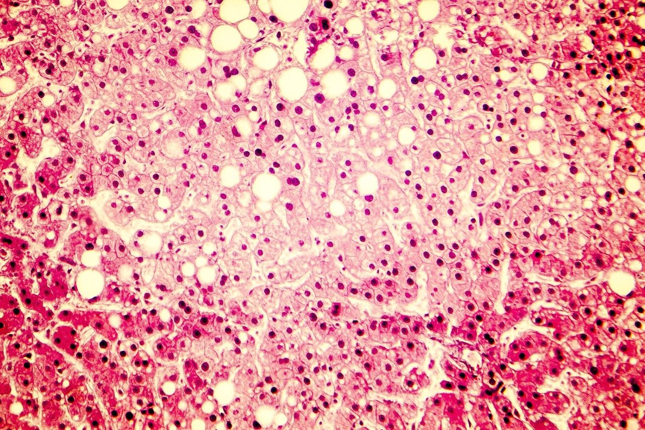
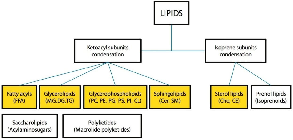
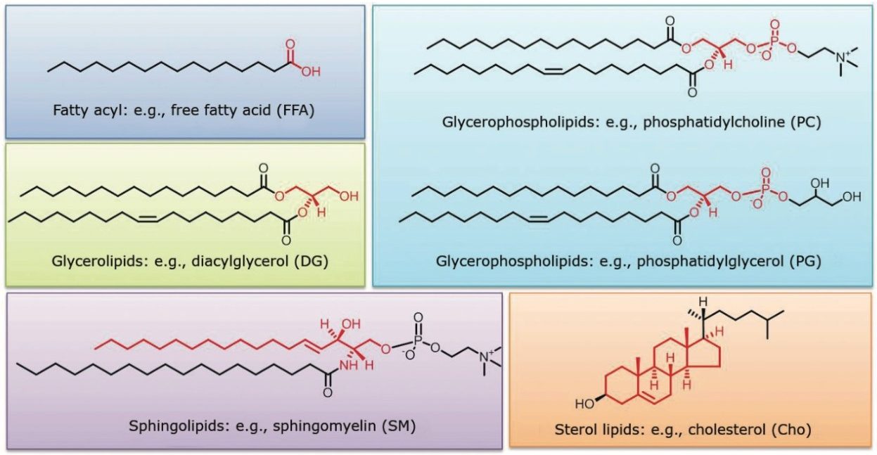
Lipids play key roles in human health. Alterations in lipid levels have been associated with the occurrence of various diseases, including cardiovascular diseases, diabetes, cancer, and neurodegenerative diseases.1 Advances in LC-MS have allowed lipids to be studied with greater sensitivity and specificity, alleviating the effects of co-eluting compounds and isobaric interference, and allowing low abundance lipids to be more readily detected.1
Conventional mass spectrometric analysis of lipids is often performed by direct infusion, or reversed-phase (RP) / normal-phase (NP) HPLC.2-5 However, each of these methods faces its own challenges.
With direct infusion, chromatographic separation of lipids is not performed prior to injection into the mass spectrometer. This method of sample introduction gives rise to ion suppression and it does not allow for separation of isobaric lipids, which can complicate the resultant analysis, necessitating deconvolution, and compromising the sensitivity of the method. In order to fully explore the lipidome, a technique of sample introduction into the mass spectrometer that minimizes these issues is needed.
NP chromatography allows separation of lipids by class but often suffers from long elution times, is difficult to handle due to the volatility and toxicity of the mobile phase, and proves challenging for ionization and introduction into mass spectrometry.6 Recent work in HILIC chromatography overcomes many of these issues.7
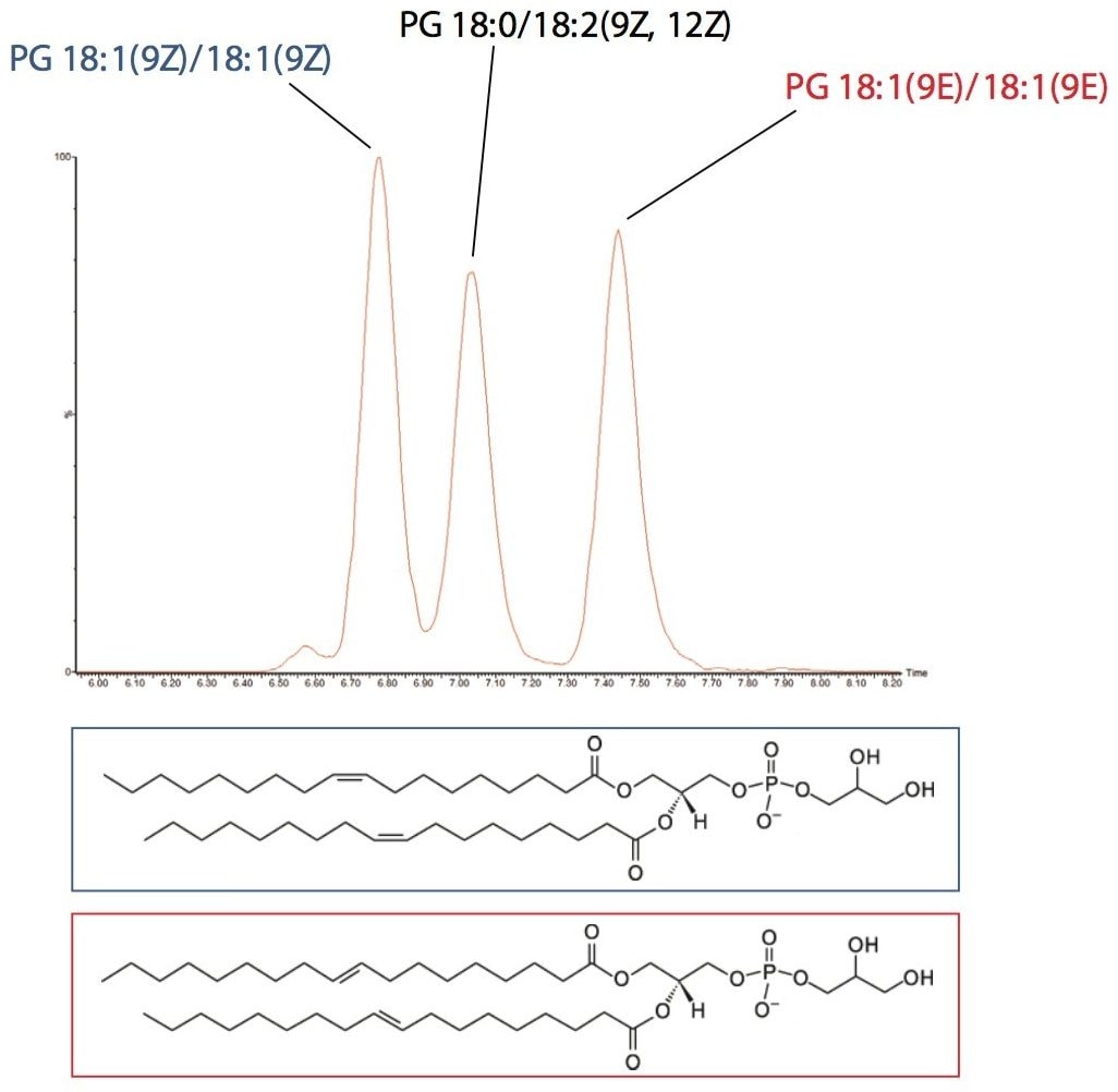
Traditional RP methods similarly suffer from extensive elution times and the quality of the resulting chromatography is relatively poor. Peak capacity and resolution are compromised in a typical analysis and it is not unusual to see peaks widths > 30 seconds,5 which ultimately results in poor sensitivity and difficulty in characterization.
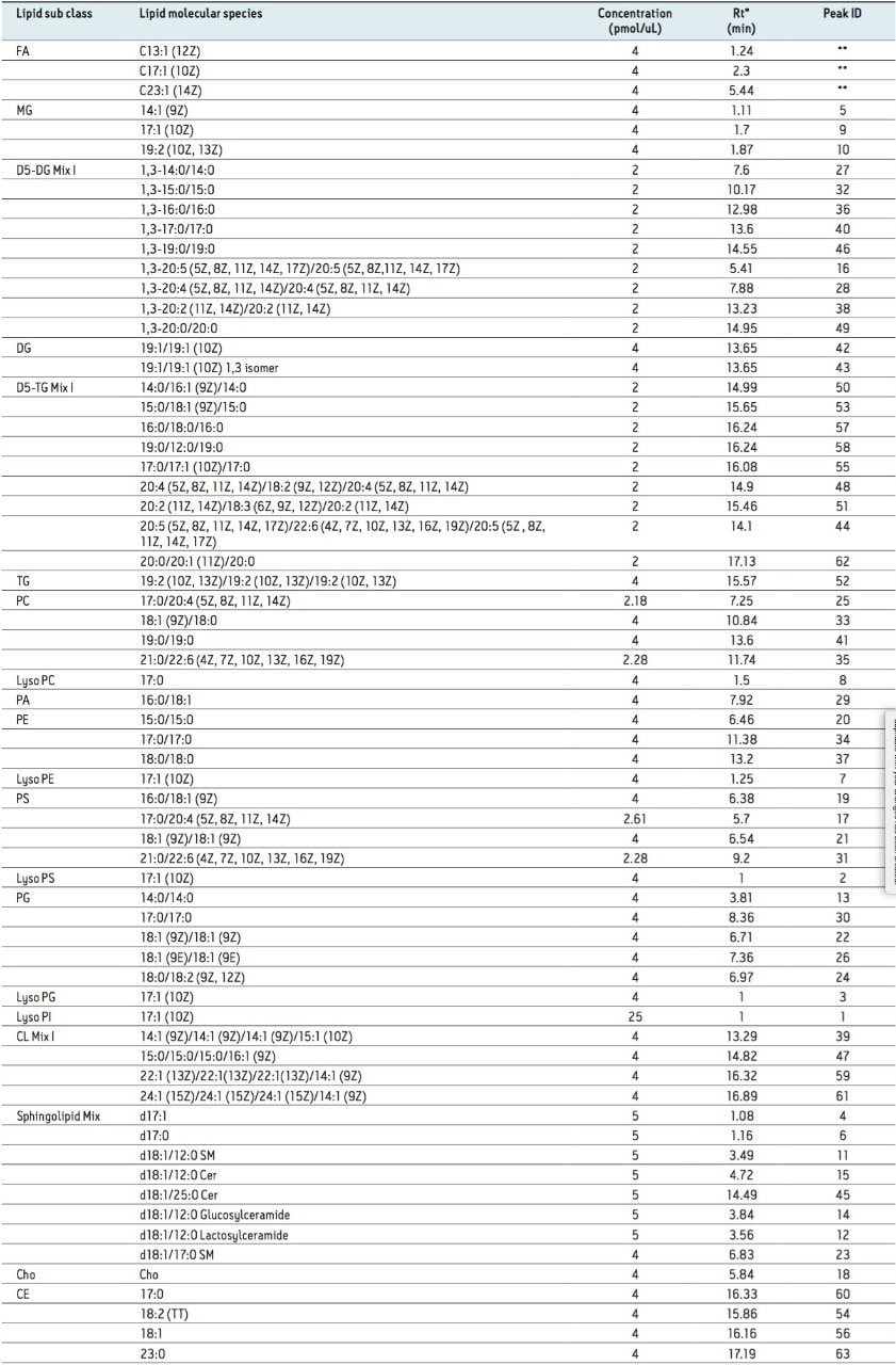
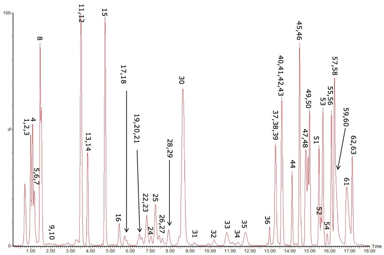
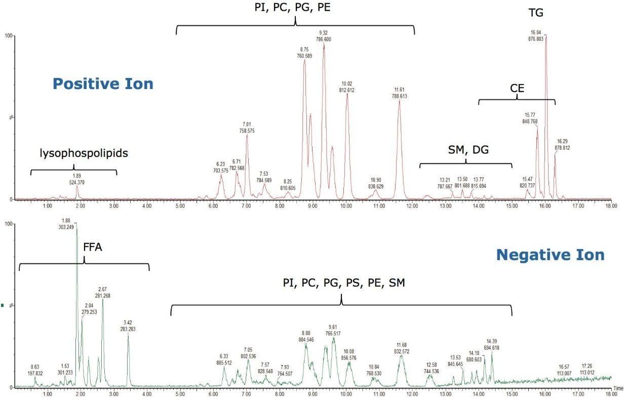
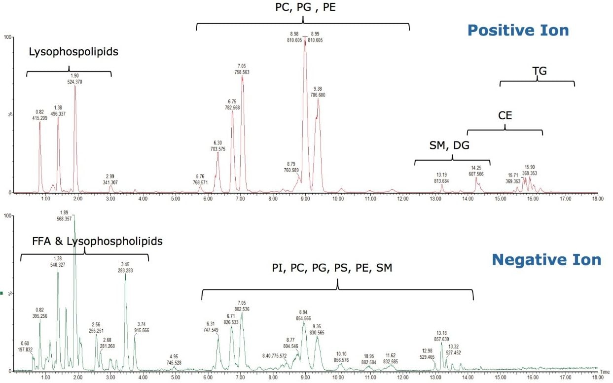
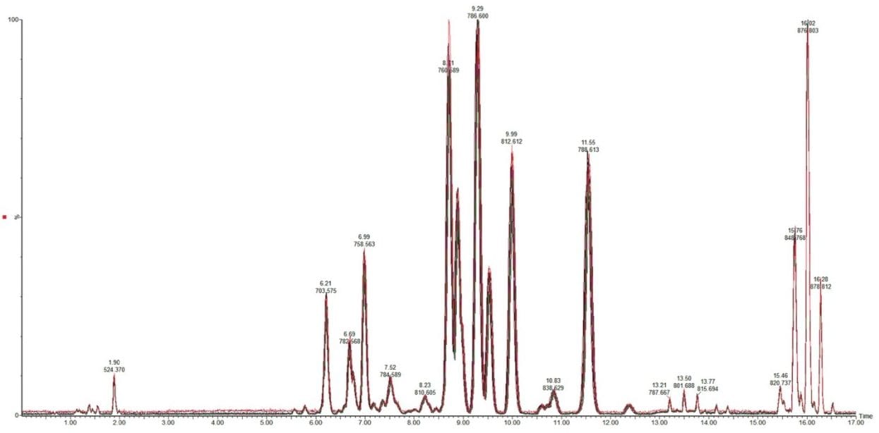
In this application note, novel RP-UPLC separations are performed using a Waters ACQUITY UPLC System with Charged Surface Hybrid (CSH) C18 chemistry. The combination of sub-2-µm particle size with an optimized liquid chromatography system and novel chemistry allows for a significantly improved RP method that maximizes the performance of these particles and is optimized for the analysis of complex lipid mixtures.