For research use only. Not for use in diagnostic procedures.
This application note describes a clinical research method utilizing Oasis PRiME HLB μElution Plate technology for the extraction of testosterone, androstenedione, and DHEAS from serum.
Testosterone, androstenedione, and dehydroepiandrosterone sulfate (DHEAS) are androgenic steroid hormones that are involved in the regulation of sexual characteristics. Analysis of these structurally similar steroid hormones by LC-MS/MS provides three levels of selectivity; sample preparation, liquid chromatography, and detection by multiple reaction monitoring (MRM) mass spectrometry. A selective sample preparation method has previously been developed which demonstrates excellent analytical sensitivity for testosterone and androstenedione using the Oasis MAX µElution SPE Plates.1 However, with the inclusion of DHEAS into the analysis, this SPE is unsuitable, as it irreversibly binds DHEAS through an anion exchange mechanism. Therefore, a more suitable sample preparation protocol for this panel of steroids is required.
Here we describe a clinical research method utilizing Oasis PRiME HLB µElution Plate technology for the extraction of testosterone, androstenedione, and DHEAS from serum, which has been automated on a Tecan Freedom Evo 100/4 Liquid Handler. Chromatographic separation was performed on an ACQUITY UPLC I-Class System using an ACQUITY UPLC HSS T3 VanGuard Pre-column and ACQUITY UPLC HSS T3 Column, followed by detection on a Xevo TQD Mass Spectrometer (Figure 1). In addition, we have evaluated External Quality Assessment (EQA) samples for testosterone, androstenedione, and DHEAS to evaluate the bias and therefore suitability of the method for analyzing testosterone, androstenedione, and DHEAS for clinical research.
System: |
ACQUITY UPLC I-Class (FTN) |
Needle: |
30 μL |
Column: |
ACQUITY UPLC HSS T3 2.1 x 50 mm, 1.8 μm (Waters P/N 186003538) |
Pre-column: |
ACQUITY UPLC HSS T3 VanGuard 2.1 x 5 mm 1.8 μm (Waters P/N 186003976) |
Mobile phase A: |
Water with 2 mM ammonium acetate + 0.1% formic acid |
Mobile phase B: |
Methanol with 2 mM ammonium acetate + 0.1% formic acid |
Needle wash solvent: |
80% methanol(aq) |
Purge solvent: |
40% methanol(aq) |
Column temp.: |
50 °C |
Injection volume: |
15 μL |
Flow rate: |
0.60 mL/min |
Gradient: |
See Table 1 |
Run time: |
4.7 minutes |
System: |
Xevo TQD |
Resolution: |
MS1 (0.75 FWHM) MS2 (0.75 FWHM) |
Acquisition mode: |
Multiple Reaction Monitoring (MRM) (see Table 2 for details) |
Polarity: |
ESI positive/negative |
Capillary: |
0.4 kV |
Source temp.: |
150 °C |
Desolvation temp.: |
450 °C |
Inter-scan delay: |
0.01 seconds |
Inter-channel delay: |
0.02 seconds |
MassLynx v4.1 Software with TargetLynx Application Manager
Testosterone, androstenedione, and DHEAS certified reference solutions and their stable labeled internal standards were purchased from Sigma Aldrich (Poole, UK). Calibrators were prepared in a surrogate matrix of MSG4000 stripped human serum purchased from Golden West Biologicals (Temecula, CA). Testosterone and androstenedione calibrators were prepared over the range of 0.17–69 nmol/L, with quality controls (QCs) at 0.52 nmol/L, 5.2 nmol/L and 35 nmol/L. DHEAS calibrators were prepared over the range of 0.14–54 µmol/L with QCs at 0.41 µmol/L, 4.1 µmol/L, and 27 µmol/L.
To convert SI units to conventional mass units divide by 3.470 for testosterone (nmol/L to ng/mL), 3.494 for androstenedione (nmol/L to ng/mL) and 2.716 for DHEAS (µmol/L to µg/mL).
Extraction was performed using a Tecan Freedom Evo 100/4 Liquid Handler. To 100 µL of sample; 25 µL of 28 nmol/L testosterone-13C3 /androstenedione-13C3, and 1.4 µmol/L DHEAS-2H5 , 200 µL methanol and 550 µL water were added. The samples were mixed after each reagent addition. Samples were centrifuged for 5 minutes at 4000 g.
An aliquot of each of the pre-treated samples (600 µL) was loaded into individual wells of the Oasis PRiME HLB µElution Plate (Waters P/N 186008052) and slowly pulled through at low vacuum (100 mbar). Consecutive washes with 200 µL of 0.1% (v/v) formic acid in 35% (v/v) methanol(aq) and 200 µL 0.1% (v/v) ammonia in 35% (v/v) methanol(aq) were performed. Analytes were eluted using 45 µL of methanol, followed by 55 µL water.
Time (min) |
Flow rate (mL/min) |
%A |
%B |
Curve |
---|---|---|---|---|
Initial |
0.6 |
55 |
45 |
Initial |
1 |
0.6 |
55 |
45 |
6 |
3.5 |
0.6 |
35 |
65 |
6 |
3.51 |
0.6 |
2 |
98 |
11 |
4 |
0.6 |
55 |
45 |
11 |
Table 1. Gradient table for the separation of testosterone, androstenedione, and DHEAS. Operating backpressure at the initial conditions was approximately 8500 psi.
No interferences were observed at the retention time of testosterone, androstenedione, and DHEAS when the analytes themselves and eight structurally related compounds were examined (11-deoxycortisol, 21-deoxycortisol, 21-hydroxyprogesterone, 17-hydroxyprogesterone, corticosterone, cortisol, DHEA, and dihydrotestosterone). The chromatographic selectivity of the column is demonstrated through the baseline resolution of testosterone and its epimer; epitestosterone (Figure 2). In addition, separation of androstenedione, 17-hydroxyprogesterone, and DHEA from testosterone is necessary due to the detection of these analytes or their isotopes in the testosterone MRM trace at concentrations of >1 µmol/L.
No system carryover was observed from high concentration samples into subsequent blank injections. High concentration samples were at 175 nmol/L for testosterone and androstenedione, and 135 µmol/L for DHEAS. A 1:5 dilution was successfully performed on the high concentration carryover sample, providing a mean accuracy of 97%, 101%, and 96% for testosterone, androstenedione, and DHEAS respectively.
Analytical sensitivity investigations reveal that the analytical sensitivity of this method would allow precise quantification (<20% RSD) at 0.17 nmol/L for testosterone and androstenedione, and at 0.14 µmol/L for DHEAS. Signal:noise (S/N) of the lowest calibration standard was >10:1 on 10 separate occasions for all analytes.
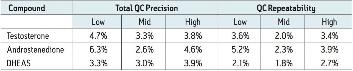
Total precision was determined by extracting and quantifying three replicates of tri-level QC material on 2 occasions per day over 5 separate days (n=30). Repeatability was assessed by analyzing three replicates at each QC level. Low, mid, and high QC concentrations were 0.52, 5.2, and 35 nmol/L for both testosterone and androstenedione. Low, mid, and high QC concentrations were 0.41, 4.1, and 27 µmol/L for DHEAS. Total precision and repeatability using the Tecan Freedom Evo 100 Liquid Handler was ≤6.3% for all analytes (Table 3).
The method was shown to be linear over the range of 0.15–76 nmol/L for testosterone, 0.15–74 nmol/L for androstenedione, and 0.13–58 µmol/L for DHEAS, when different ratios of high and low concentration pools of the analytes were combined and analyzed. In addition, calibration lines in spiked serum were linear with coefficient of determinations (r2) >0.998 on 10 separate occasions for all analytes.
Matrix effect investigations were performed using individual donor serum samples. The endogenous peak areas were separately quantified. Post-spiked samples were adjusted using the mean endogenous peak area to enable comparison to solvent spiked samples. The matrix factor calculated is shown in Table 4. Normalized matrix factor calculations, based on the analyte:internal standard response ratio produced similar values to peak area matrix factor.
Samples were selected (n=50) for comparison against an independently developed LC-MS/MS method for testosterone, androstenedione, and DHEAS. Comparison data was processed using Analyse-it v2.3. Altman-Bland agreement demonstrated a mean bias of 5.0%, -3.3%, and -6.3% for testosterone, androstenedione, and DHEAS, respectively.
EQA samples were analyzed for testosterone (n=30), androstenedione and DHEAS (n=50). The data obtained was compared to the mass spectrometry mean for the samples and Deming regression was performed. The correlation for testosterone, androstenedione, and DHEAS can be seen in Table 5 showing the excellent agreement with the EQA scheme MS mean. Proportional and constant bias was observed for DHEAS when all samples were analysed (0–40 μmol/L), however, the bias over the range 10.2–14.9 μmol/L was ≤6.9%. Altman-Bland agreement demonstrated a mean bias of -0.50%, 0.4%, and 5.8% for testosterone, androstenedione, and DHEAS, respectively.
An analytically sensitive and selective clinical research method has been developed for the analysis of testosterone, androstenedione, and DHEAS in serum.
Using only 100 µL sample volume, this method provides sufficient analytical sensitivity to analyze low physiological levels of testosterone, androstenedione, and DHEAS. Automation of the analytical method in combination with sample tracking capabilities improves laboratory workflow and reduces sample handling, which alleviates the potential for operator error.
Professor Brian Keevil and his colleagues at the Department of Clinical Biochemistry, University Hospital of South Manchester, Wythenshawe, UK, are thanked for the provision of anonymized serum samples for the analysis.
720005554, December 2015