Rapid Automated Antibody Purification Using Protein A-Coupled Magnetic Beads With Andrew+
For research use only. Not for use in diagnostic procedures.
Abstract
Monoclonal antibodies remain one of the rapid growing classes of biopharmaceuticals.1 High-throughput small-scale analytical platforms are increasingly sought after within general research and to aid in the development of biopharmaceuticals when using miniaturized bioreactors.1 In this regard, affinity purification is one of the most widely employed purification strategies prior to analysis because high purity antibody samples are often required for further analysis.2 As an easy-to-use separation medium, magnetic beads streamline the purification process without the need to use complex instrumentation.2
Currently, in the field of magnetic separation, many existing automated protocols still require several manual intervention steps.3 A fully automated antibody purification protocol will increase throughput, save time, and increase the efficiency of analytical workflow in biotherapeutics.
Here, we present a rapid and fully automated antibody purification protocol that can isolate target antibody from cell culture media using Andrew+ pipetting robot. The protocol can be easily adjusted to accommodate samples with either high or low titer to yield purified antibodies at the desired concentration. In addition, samples of low titer can be concentrated to produce purified antibody at a higher concentration.
The rapid protocol is fully automated, reproducible, robust, and is highly adaptable for further analysis.
Benefits
- Fully automated Protein A magnetic beads purification protocol with Andrew+
- Rapid purification protocol (35 min for eight samples)
- Greater than 75% recovery for antibody loads of 20 µg to 180 µg and media sample titers ranging from 0.2 μg/μL to 1.5 μg/μL
Introduction
During process development, manufacturing, and quality control (QC) of therapeutic antibodies, purification is routinely carried out.4 Affinity purification using Protein A is a widely used purification strategy due to the selectivity and high binding affinity of Protein A to IgG antibodies.1 Similarly, magnetic beads are a commonly used medium for purification due to their ease of use.1 Protein A-coupled magnetic beads combine the benefits of high selectivity of protein A and effective removal of the beads from the purified sample.1
Existing automated protocols using the magnetic separation method require several manual intervention steps such as adding magnetic beads to a 96-well plate, transferring the plate, adding buffers to the plates during washing and elution steps, and transferring the purified samples to a fresh plate.3
Here, we describe a fully automated antibody purification protocol using the Andrew+ pipetting robot (p/n: 176850100). The robot uses dominos, a microplate grabber (p/n: 186009776), and the connected devices such as 96-PCR Plate Magnet+ (p/n: 186009956), and Microplate Shaker+ (p/n: 186009594) and requires minimal manual intervention. The automation capabilities of Andrew+ allow for the flexibility of processing samples using either a single-channel or 8-channel pipetting depending on the throughput needs.
Experimental
Sample Description
Conditioned cell culture media samples, with CHO cells removed, were provided by a collaborator. Clarified samples were used without further centrifugation. Other reagents and chemicals used were shown in the table below.
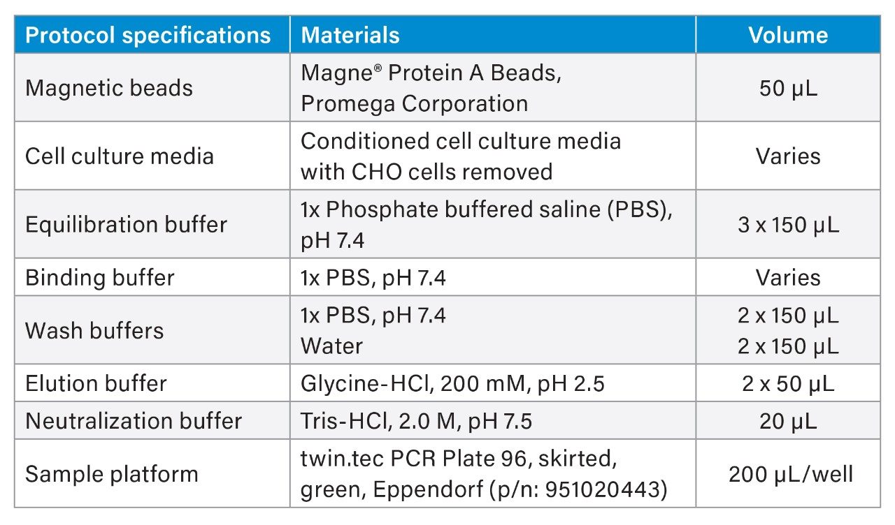 Table 1. Reagents used for antibody purification protocol.
Method Conditions
|
LC system: |
ACQUITY UPLC H-Class Bio System |
|
Detection: |
TUV detector 220 nm and 280 nm |
|
Sample collection: |
twin.tec PCR Plate 96, skirted, green, Eppendorf, (p/n: 951020443) |
|
Column: |
ACQUITY UPLC Protein BEH SEC, 200 Å, 1.7 µm, 4.6 x 150 mm (p/n: 186005225) |
|
Column temp.: |
Off |
|
Sample temp.: |
4 °C |
|
Needle placement: |
5.0 mm (default is 2.0 mm) |
|
Injection volume: |
2 µL |
|
Flow rate: |
0.4 mL/min |
|
Run time: |
7 min |
|
Mobile phase: |
100 mM ammonium acetate, pH 5.25 (acetic acid) |
|
Gradient: |
Isocratic |
Data Management
|
Chromatography software: |
Empower 3 |
Instrumentation
To prevent a magnetic bead from accidentally getting into the LC system, two accomodations were put in place in the procedure. A magnet (96-Well Deep Bar Magnet Plate, Permagen, p/n: MSPU650 ) was placed under the 96-well PCR plate in the sample manager to fully capture any rogue beads in the final sample. The modified plate parameters for LC are shown in Figure 1. The needle placement in the LC method was changed to 5.0 mm from the default value of 2.0 mm to accommodate the change.
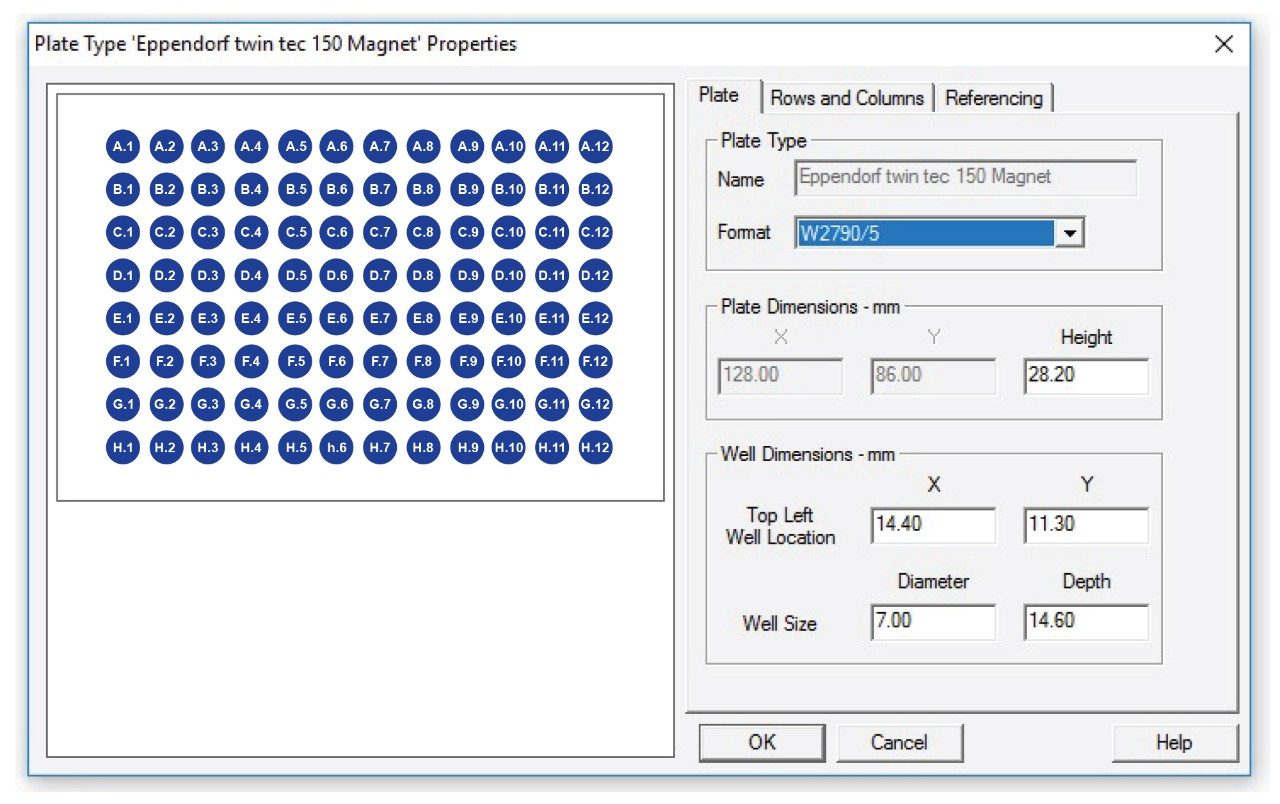 Figure 1. Plate parameters used in Empower to accommodate the 96-well PCR plate with a bar magnet underneath.
Results and Discussion
The general principle behind the antibody purification with the magnetic beads is illustrated in Figure 2.5 Briefly, samples are first incubated with protein A-coupled magnetic beads in a binding buffer for a certain amount of time with gentle mixing. Then, impurities are washed away, and bound antibodies are eluted from the beads using an acidic elution buffer.
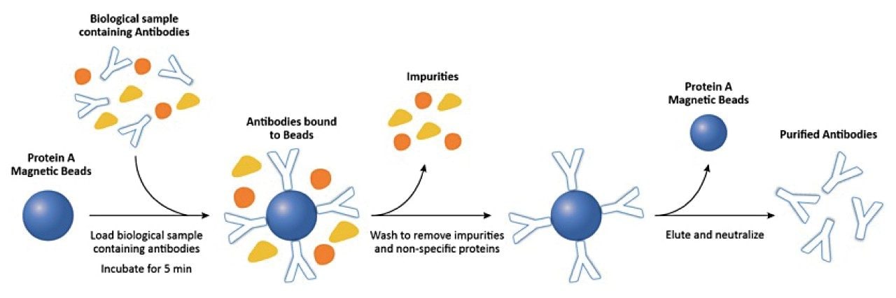 Figure 2. General protocol for antibody purification using Protein A-coupled magnetic beads.
Rapid automated antibody purification protocol is essential to the development of biotherapeutics
The general protocol is relatively simple yet consists of several equilibration/washing steps, and a manual execution of a routine protocol for multiple samples can be laborious and time consuming. Using our fully automated protocol, users can perform the antibody purification without any manual intervention step, increasing sample throughput. The flow diagram describing the steps performed during the automation experiments are shown in Figure 3. Three procedures are presented here to accommodate the varying titers and loads of antibody (low ≤50 µg, standard ≤75 µg, high >75 µg) in the cell culture media. Low load protocol has additional steps to load the samples multiple times so that more concentrated samples are eluted at the end. If the purified antibody of higher titer sample is not required for further analysis, the standard protocol (load 50 µg, 0.5 mg/mL) can also be used for low titer samples. High load protocol has a longer incubation time of 10 min, instead of the standard incubation time of 5 min. Samples of high titer may also be diluted with the binding buffer (1x PBS, pH 7.4) before loading, if necessary.
Automation capability of Andrew+
In the field of magnetic separation and protein purification, Andrew+ provides a streamlined fully automated protocol. In comparison, other magnetic bead handlers provide a semi-automated protocol that requires one to five manual intervention steps for a similar procedure.3
Rapid automated antibody purification protocol improves sample throughput
The rapid automated protocol presented here needs minimal manual interventions during the purification process and the total execution time for eight samples is 35 min. In comparison, the total run time needed to complete the protein purification on other similar device is one hour, 20 min when using the same type of magnetic beads from Promega.6 Therefore, Andrew+ can effectively streamline the antibody purification with magnetic beads using the connected devices as shown in Figure 4.
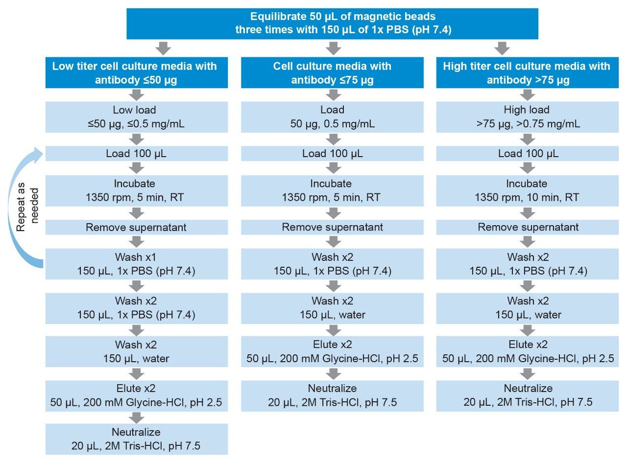 Figure 3. Flow diagram of automated antibody purification procedures. Three procedures were presented here to accommodate varying titers and loads of antibody (low ≤50 µg , standard ≤75 µg, high >75 µg) in cell culture media. RT: room temperature.
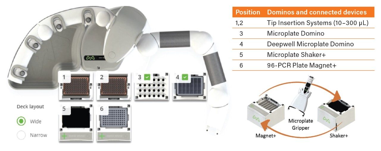 Figure 4. Andrew+ Dominos and Connected Devices configuration for the rapid automated antibody purification eight-sample protocol. For a fully automated protocol, Andrew+ requires two connected devices (Magnet+ and Shaker+) and a microplate gripper or a microplate grabber. The execution time for eight samples is 35 min.
Automation development of antibody purification protocol
Automation development included the adjustments of pipetting settings, tip position, pipetting technique, and shaker speed to maximize recovery.
Pipetting settings/Handling liquid viscosity
Due to the low viscosity and high volatility of 20% ethanol solution, in which the magnetic beads are suspended, the pipette settings were adjusted for low viscosity mode with faster than normal speed of aspiration and dispensing. This will ensure to dispense a similar number of beads into all the wells. If using a different magnetic bead supplier, these settings may need to be adjusted.
For cell culture media samples, the pipette settings were adjusted to pipette each sample using high viscosity mode to assure slow aspiration and dispensing of the samples without vigorous stirring, which can introduce air bubbles to the protein solution.
Tip position
Regarding the pipette tip position, “With respect to liquid” option was used for all the equilibrating steps so as not to aspirate and discard the magnetic beads before purification. “With respect of bottom” option was used for all other steps to remove all the binding buffer completely before elution.
Pipetting technique/Mixing
Multiple aspirations are used to ensure thorough mixing and distribution of samples. Mixing the samples several times at the source will ensure the equal distribution of magnetic beads. Magnetic beads tend to settle, and the solution need to be mixed right before aspirating into the individual wells. Thus, repetitive mode of pipetting must be avoided. It is recommended to have 0.5–1.5 mL of magnetic beads slurry in the source tube to enable thorough mixing of the slurry. For 0.5–1.5 mL slurry, thorough mixing was obtained by using mixing volume of 300 µL.
Cell culture media samples also need to be mixed before aspiration and thus, a similar pipetting technique (aspirate three times at the source before transfer) was used.
Shaker speed and time
It is imperative that during the binding steps that the beads must remain in suspension for maximum binding, which is accomplished using the Microplate Shaker+ (Figure 4). The optimum speed and time were found to be 1350 rpm and 5 min. Sample shaker speed and time may need to be adjusted, if a different magnetic bead supplier is used. However, it is important that the shaking is not too vigorous, which can result in excessive sample foaming.
Determination of antibody recovery
Antibody recoveries from protein A purifications were determined based on the SEC peak area of the mAb monomer as measured for both the conditioned media and Protein A purified samples. While the co-elution of lower abundance host-cell proteins with the mAb in the SEC separation of the conditioned media is possible, the co-elution was considered to have a minimal effect on the results and would only result in an underestimation of antibody recovery for the Protein A purification. Figure 5 shows the size exclusion chromatograms of media before purification (Figure 5A, green trace), after purification (Figure 5C, red trace), and supernatant (Figure 5B, orange trace). Supernatant consists of the components in the media other than the antibody and it is the solution left over after the antibody is bound to the Protein A on the magnetic beads.
%Recovery = 100 * (Adjusted AreaPurified /AreaProteinA Load )
Adjusted AreaPurified# = AreaPurified* (Total VolumePurified)/(Total VolumeProteinA Load )
#The peak area of the eluted protein (AreaPurified) was adjusted as shown in the second equation because the eluted volume of protein (Total VolumePurified) is 120 µL whereas the loaded volume of protein (Total VolumeProteinA Load) is only 100 µL.
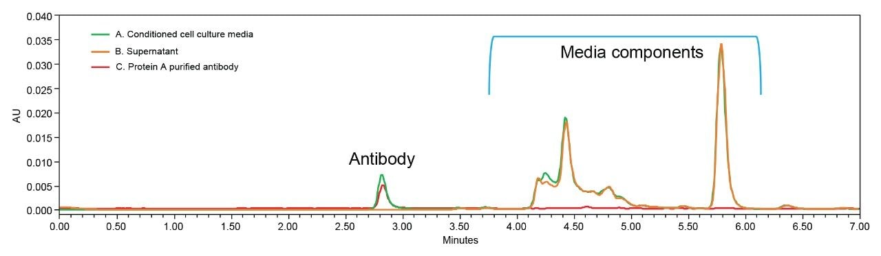 Figure 5. Size exclusion chromatograms of cell culture media samples before purification (A, green trace), after purification (C, red trace), and supernatant (B, orange trace).
Rapid automated antibody purification protocol is reproducible and robust
Using the rapid automated protocol, Andrew+ can achieve a high recovery of antibody with the recovery values generally greater than 75% for samples with a titer of approximately ≤0.75 mg/mL with a reliable reproducibility (%RSD ≤10%). Recovery obtained from the manual execution of the procedure and the automated procedure are comparable as shown in Figure 6.
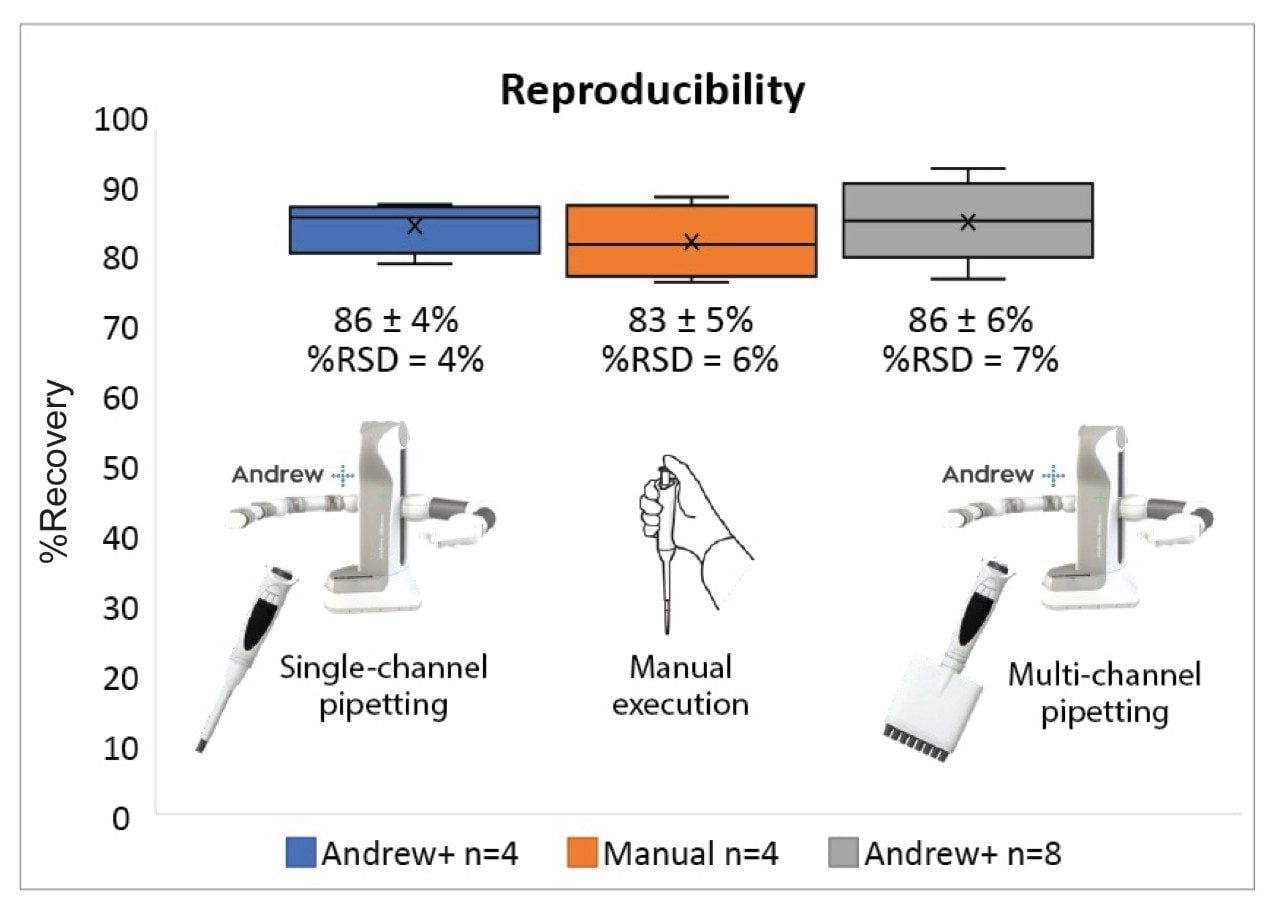 Figure 6. Comparison of the recoveries of antibody obtained from the manual execution (n=4) and the Andrew+ Pipetting Robot (n=4 and n=8). Markers (x) represent the average values.
The robustness of the automated protocol was demonstrated by varying the amount of Protein A magnetic beads used for the capture step (Figure 7A) and the pH of the elution buffer (Figure 7B). The use of magnetic beads requires pipetting the beads from a homogenous slurry. To evaluate the impact on antibody recovery by the number of beads used, the standard amount of the slurry used (50 μL) was varied by ±10%. These experiments with variations still produced satisfactory recovery of antibody (>50%, Figure 7A).
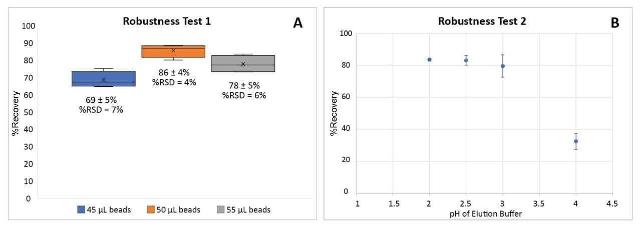 Figure 7. Robustness assessment of the automated protocol. Comparison of the recovery of antibody (A) with three different amounts of beads (45 μL, 50 μL [standard], 55 μL), n=4 and (B) with four different buffered pH levels (pH = 2, 2.5 [standard], 3, 4), n=2. Markers (x) represent the average values.
Another variable that may have a significant impact on antibody recovery is the pH of the elution buffer. Our standard protocol uses Glycine-HCl (0.2 M) at pH 2.5 as the elution buffer. To test the robustness, elution buffers with a pH ranging from 2 to 4 were used for elution (Figure 7B). Recovery was not affected when the pH of the elution buffer was between 2 and 3. However, a low recovery was observed with the elution buffer with pH 4. Therefore, the purification method is robust between pH 2 and 3.
Rapid automated protocol can be used for both low and high titer samples
For samples with low titer (≤50 μg), multiple loadings can be performed to achieve a highly concentrated purified antibody. Three 100 μL loadings (5 min each) of cell culture media with a titer of 0.6 μg/μL (total of 180 μg) with an alternating washing step, yielded a comparable recovery of antibody (80%) to that of single loadings of media samples (Figure 8).
While the standard incubation time of 5 min was adequate for low titer samples, for samples with high titer (>75 μg), a longer incubation time of 10 min was necessary to obtain a high recovery. The purification of samples with a media load of 150 μg resulted in 65% recovery with a 5 min incubation. Prolonging the incubation time from 5 min to 10 min increased the yield from 65% to 87%, as shown in Figure 8. Alternatively, the samples of high titer can also be diluted with the binding buffer prior to loading.
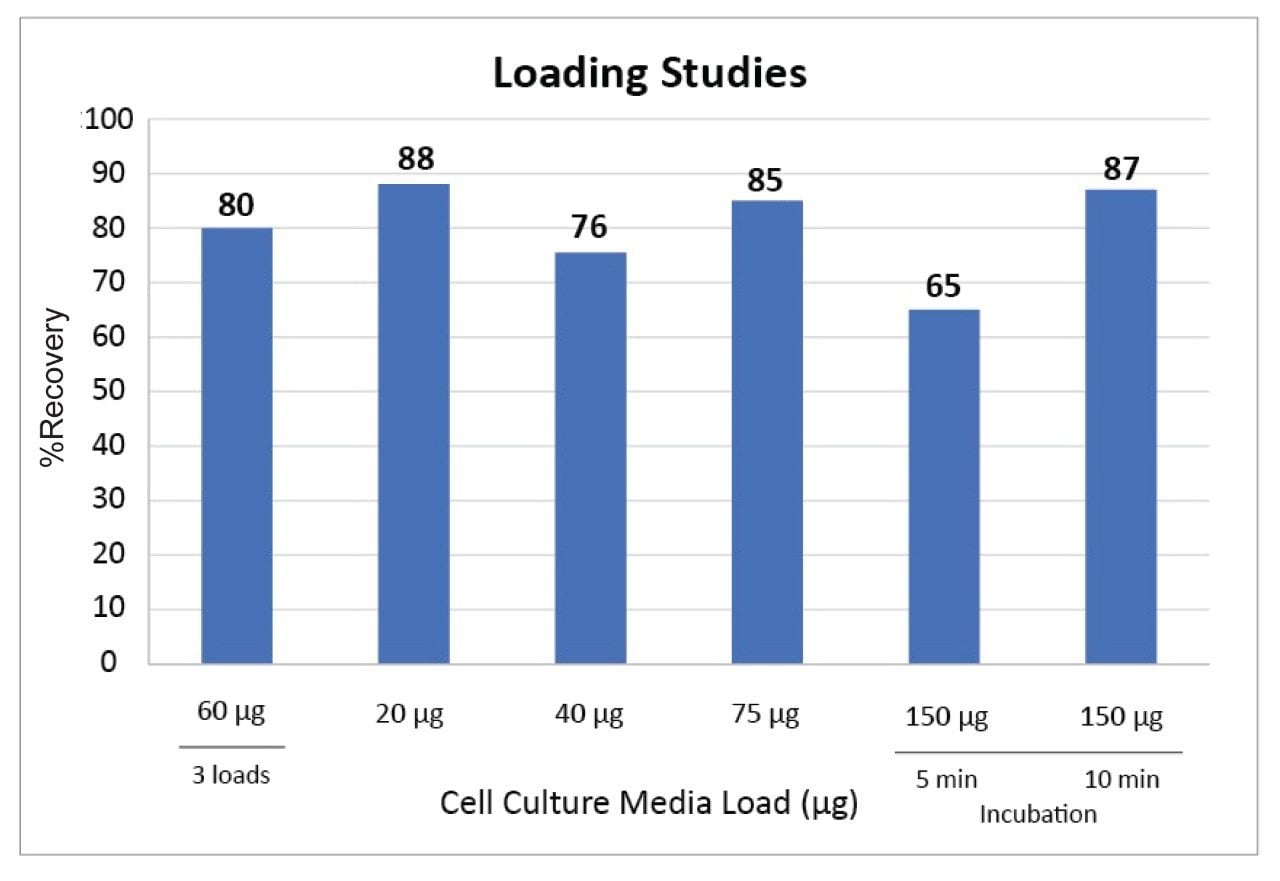 Figure 8. Antibody recovery from cell culture media samples with various loads (20 μg, 40 μg, 75 μg, 150 μg, 180 μg) of antibody. Multiple loadings were obtained by loading 60 μg three times with an intermediate washing step (n=2). Loading volumes were 100 μL. Titers for media samples ranged from 0.2 μg/μL to 1.5 μg/μL.
Conclusion
The rapid antibody purification protocol is reproducible, robust, and can extract antibodies in a high yield (>75% recovery) from cell culture media. The protocol is fully automated, requires no manual intervention, and increases throughput. The protocol can be easily adjusted for both high and low titer samples to yield the purified antibodies at the desired concentration. In addition, low titer samples can be concentrated to obtain pure antibody at a higher concentration.
References
- Choe, W.; Durgannavar, T. A.; Chung, S. J. Fc-Binding Ligands of Immunoglobulin G: An Overview of High Affinity Proteins and Peptides. Materials 2016, 9, 994.
- Brechmann, N. A.; Eriksson, P.; Eriksson, K.; Oscarsson, S.; Buijs, J.; Shokri, A.; Hjalm, G.; Chotteau, V. Pilot-Scale Process for Magnetic Bead Purification of Antibodies Directly From Non-Clarified CHO Cell Culture. Biotechnol. Prog. 2019 Jan, 35(3), 1–10.
- Kuhn, E.; Fabbami, L.; Heuvel, Z. V. D.; Murphy, S.; Jaffe, J. D.; Carr, S. A. Automation of the Multiplexed Peptide Immune-MRM-MS Workflow on Bravo AssayMAP Platform. Broad Institute, Cambridge, MA; Agilent Technologies, Santa Clara, CA.
- GE Healthcare. Antibody Purification Handbook.
- Magne Protein A Beads and Magne Protein G Beads for Antibody Purification. Promega Corporation: Madison, WI, 2015.
- Bratz, M.; Godat, B.; Wieczorek, D.; Nath, N. A Robist High-Throughput Method for Antibody Purification using Magnetic Beads on the KingFisher Flex Platform. Promega Corporation: Madison, WI, 2016.
- Li, Feng; Vijayasankaran, N.; Shen, A.; Kiss, R. l.; Amanullah, A. Cell Culture Processes for Monoclonal Antibody Production. mAbs. 2010 Sept, 2(5), 466–477.
Acknowledgments
We thank our collaborators for their generous supply of cell culture media.
720007343, August 2021