• Application Note

GlycoWorks RapiFluor-MS Automation Using the Andrew+ Pipetting Robot

GlycoWorks RapiFluor-MS Automation Using the Andrew+ Pipetting Robot

  • Corey E. Reed
  • Stephan M. Koza
  • Steven Calciano
  • Waters Corporation

This is an Application Brief and does not contain a detailed Experimental section.

Abstract

Using the Andrew+ pipetting robot from Andrew Alliance - A Waters Company, the GlycoWorks RapiFluor-MS N-Glycan Labeling Kit was automated and verified for performance. Verification requirements included comparability to manually performed preparations of the same sample number with ≤25% deviation in total area and ≤5% deviation in relative area. During verification, it was evident that protocol optimization was required due to changes in consumables causing non-specific sample losses of 45% on average during the automated protocol. Changes were made to mitigate this sample loss, and ultimately the automated protocol showed 88% recovery compared to the manual protocol. Throughout the optimization process relative areas remained highly reproducible, and the final protocol showed no relative area deviation >2.6% from the manual procedure.

Benefits

  • Ease-of-use and time savings
  • Cost savings through increased efficiency and failure reduction
  • Cost efficient robotics solution
  • Collaborative automation
  • Rapid N-glycan labeling
  • Reduced analyst stress
  • Removal of analytical bottleneck
  • Automated method transferability

Introduction

Glycosylation is routinely monitored as a critical quality attribute (CQA) during biopharmaceutical drug development as it is a measure of manufacturing condition uniformity, product efficacy, and product safety.1,2 Traditional released N-glycan labeling methods can take multiple hours or days to complete. Additionally, traditional labels often lack stability, and do not offer strong fluorescence and mass spectrometric sensitivity at the same time, limiting the research laboratory to one detection method or the other.

Waters’ introduction of the GlycoWorks RapiFluor-MS N-Glycan Labeling Kit and associated protocol provided a well-documented decrease in the time required to achieve unbiased labeling while simultaneously increasing fluorescence and mass spectrometric detection.3 The labeling workflow along with HILIC cleanup and sample collection could be completed in under an hour depending on sample number, and the simplicity of the method leant itself to automation.4 The primary benefits of automating this procedure are reducing the time spent by the analyst to prepare samples, reducing training and documentation burdens, and reducing potential errors due to pipetting monotony. With this in mind, the GlycoWorks RapiFluor-MS Kit was automated on the Andrew Alliance pipetting robot, Andrew, in 2018.5

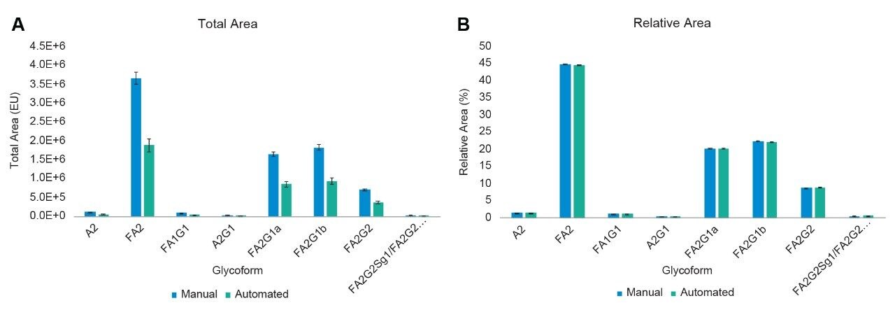
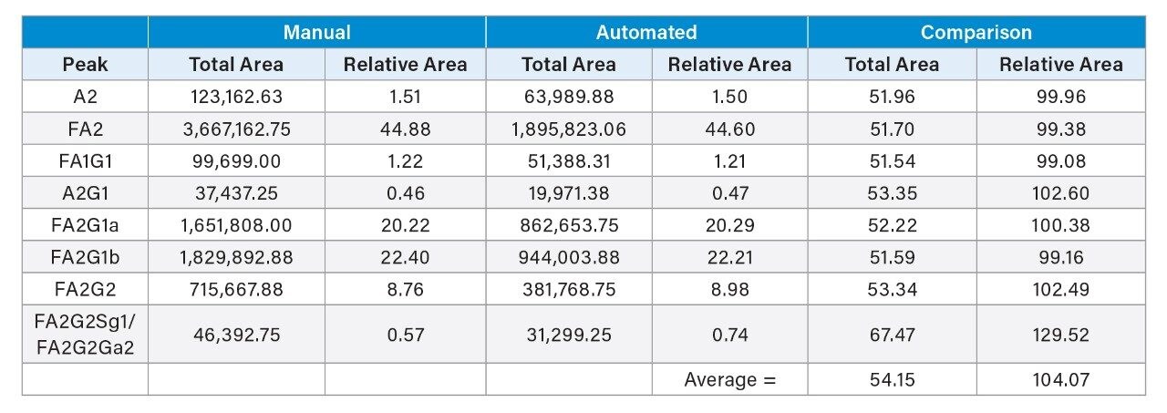
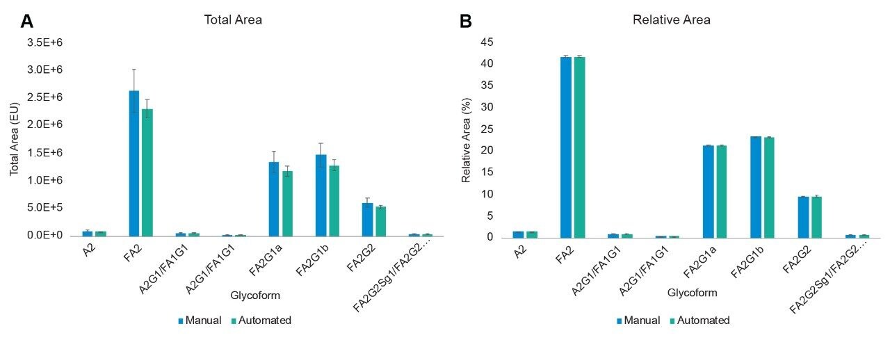
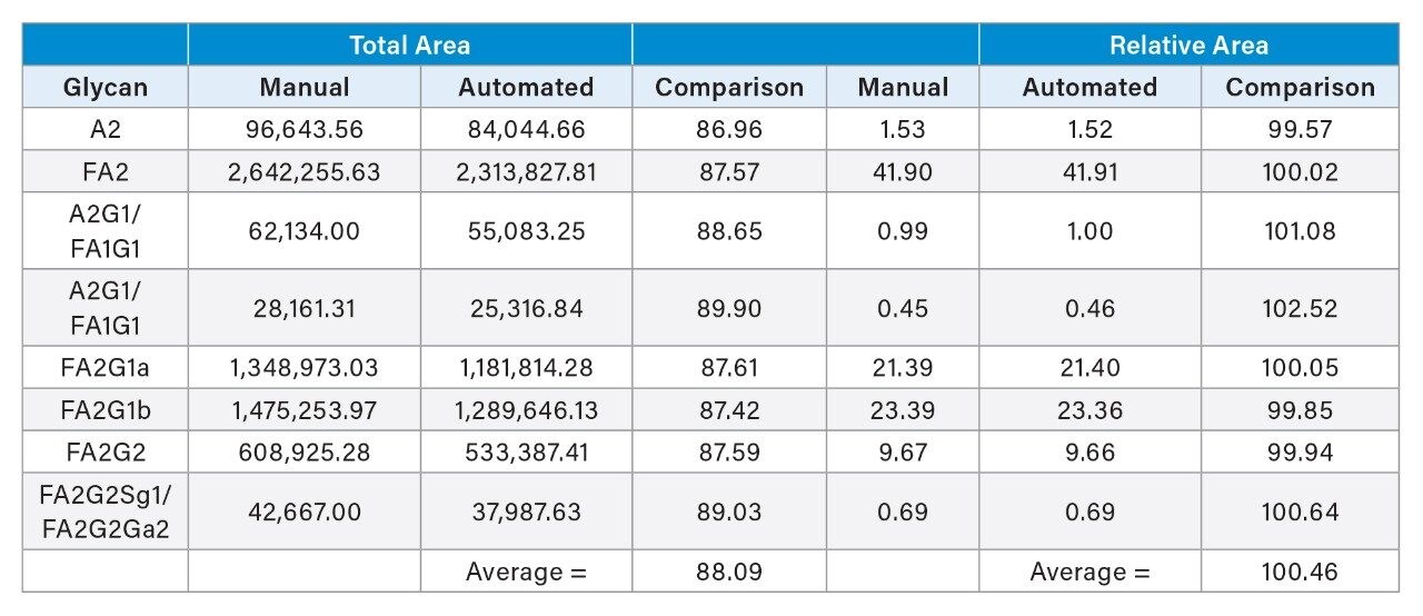
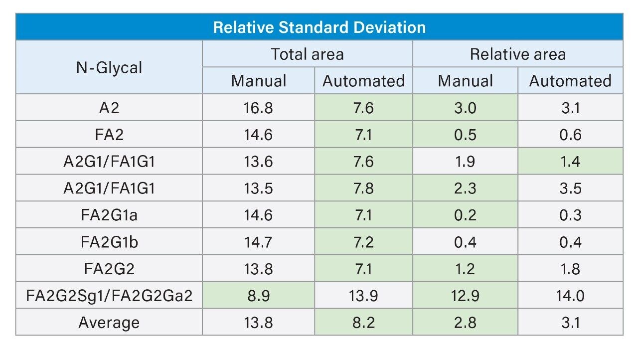
The automated protocol underwent several rounds of optimization to ensure complete release and labeling of monoclonal antibody (mAb) N-glycans comparable to that seen when performing the protocol manually. The final protocol achieved relative standard deviations of 9-19% for major and minor glycoforms released from a murine mAb standard compared to the same sample prepared by a manual user.

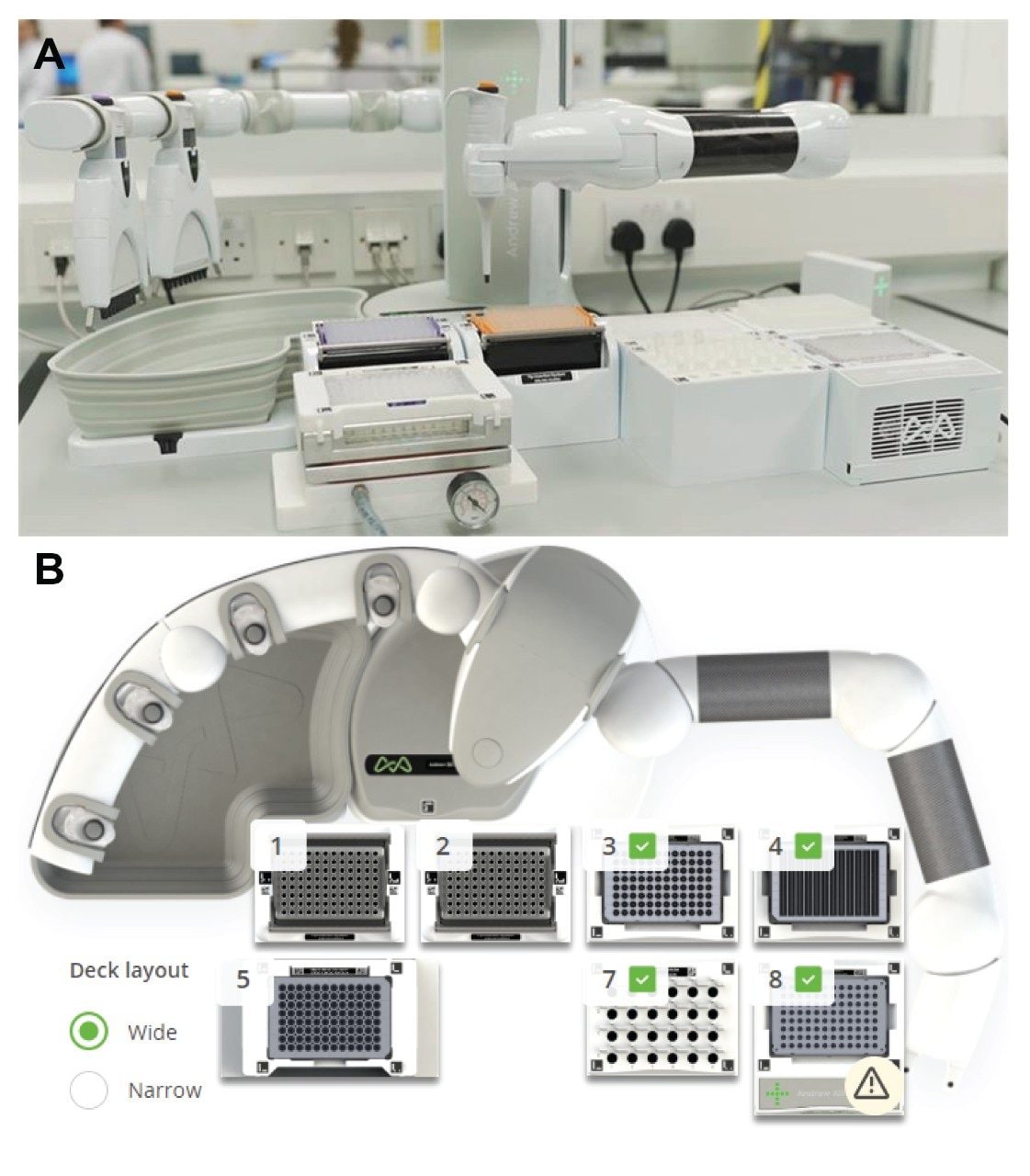
In 2019, Andrew Alliance released an updated version of the pipetting robot, named Andrew+ (Figure 1). This system features web-based connected devices and an improved robotic arm compatible with single and multi-channel electronic pipettes, leading to time savings as well as the ability to handle higher sample loads. In this application brief, the process of optimization for the 8-sample GlycoWorks RapiFluor-MS released N-glycan protocol is detailed.

Figure 1. A) Picture of the Andrew+ setup for an 8-sample GlycoWorks RapiFluor-MS protocol. B) OneLab top-down view of the Andrew+ setup for the 8-sample GlycoWorks RapiFluor-MS protocol. Dominos on the automation deck space include: (1) Tip Insertion System Domino fitted with 50 – 1200 µL Optifit tips; (2) Tip Insertion System Domino fitted with 10 – 300 µL Optifit tips; (3) Storage Plate Domino fitted with QuanRecovery with MaxPeak HPS 700 µL, 96-well plate; (4) Deepwell Microplate Domino fitted with Axygen 12-well reservoir with 12-channel trough; (5) Microelution Plate Vacuum+ Domino fitted with GlycoWorks HILIC µElution plate; (7) Microtube Domino fitted with Fisherbrand premium 1.5 mL conical microtubes for PNGase F Enzyme, RapiFluor-MS Labeling Reagent, and RapiGest-SF Surfactant; (8) 96-PCR Plate Peltier+ Domino fitted with an Eppendorf twin.tec 96-well skirted LoBind PCR plate containing samples for analysis.

Results and Discussion

Several consumable changes were implemented during development of the automated protocol on Andrew+. This was to ensure compatibility with SBS/SLAS microplate specifications6 to integrate standardized labware and connected devices for heating and vacuum. During a manual protocol, 1.2 mL sample reaction tubes are used for protein denaturation, de-glycosylation, and labeling with heat blocks pre-set at the desired temperatures. The automated protocol leverages a high speed SBS format Peltier device, however it was not amenable to the 1.2 mL tubes of the manual workflow. Therefore, 200 µL 96-well Eppendorf Lo-Bind PCR plates were implemented for the reaction steps. The plate format is preferred to individual tubes for the automation protocol as it is compatible with a wide array of connected devices and the 8-channel pipette. However, the smaller 200 µL volume of the Eppendorf plate forced changes during a critical point in the protocol.

Following the labeling step of the protocol, the 40 µL reaction volume is diluted with 360 µL acetonitrile, bringing the total reaction volume to 400 µL, which is not compatible with the 200 µL, 96-well PCR plate. As a workaround, a 1 mL 96-well plate was used to dilute the reaction volume to 400 µL before sample addition to the HILIC µElution plate. The diluted volume is then transferred to the HILIC plate for cleanup before analysis. The analytical method used can be found in the GlycoWorks RapiFluor-MS N-Glycan Kit care and use manual (p/n: 715004793EN). Figure 2 shows the results of the pre-optimized automated protocol compared to an experienced manual user.

Figure 2. Eight-sample manual and automated sample preparations of Intact mAb Mass Check Standard (p/n: 186006552) murine IgG1 protein executed on the same day, analyzed in the same sample set. Four of the eight manual samples were not analyzed for expediency. All eight automated samples were analyzed to ensure there were no complete failures. Samples were averaged and error bars are the standard deviation of all samples. A) Total area comparison across all eight glycans monitored from the chromatographic profile. B) Relative area comparison for the same eight glycan peaks.

This automated protocol was not comparable to a manual user. Total area comparison showed that the automated preparation resulted in the recovery of 54% of glycans on average over the 8 glycans monitored as compared to the manual procedure (Table 1). It is important to note that the sample loss seen is unbiased, as relative areas remain constant between the automated and manual sample preparations. This result fell well outside the acceptance criteria of the automated method being within 25% of the manual method for total area.

Table 1. Total area counts in emission units (EU) and relative area counts (%) for the automated and manual sample data shown in Figure 2 with a comparison of automated to manual samples.

Troubleshooting of the protocol was necessary to determine the source of sample loss. After eliminating the reaction steps (denaturation, deglycosylation, and RapiFluor-MS labeling) and sample cleanup as potential points of significant analyte loss, it was deduced that the primary point of loss was the dilution of sample in the 1 mL 96-well plate. Analyte recovery during this dilution step could not be adequately improved by altering the procedure or labware.

However, when the storage location was removed from the protocol and samples were mixed directly in the µElution plate, the automated sample recovery increased significantly (Figure 3). The final automated 8-sample GlycoWorks RapiFluor-MS protocol pre-loads the HILIC µElution plate with 200 µL of acetonitrile, then dilutes the reaction up to 200 µL with 160 µL acetonitrile which is transferred to the HILIC plate and mixed to the final 400 µL volume. This protocol shows 88% recovery compared to an experienced manual user. As with all other tests, the relative areas were highly comparable between the manual and automated preparations, including with low level glycans (Table 2). It is important to note that intra-preparation relative standard deviations indicate that the total area reproducibility of the automated solution is superior to a manual user (Table 3). The relative area’s relative standard deviations are comparable between the preparation methods.

Figure 3. Final GlycoWorks RapiFluor-MS 8-sample protocol. Data is cumulative from two 8-sample protocols for both manual and automated prepartions. N = 16, in all cases. A) Total area comparison across all eight glycans monitored from the chromatographic profile. B) Relative area comparison for the same eight glycan peaks.
Table 2. Total area counts in emission units (EU) and relative area counts (%) for the final automated GlycoWorks protocol sample data shown in Figure 3 with a comparison of automated to manual samples.
Table 3. Relative standard deviation comparison of the manual and automated 8-sample GlycoWorks preparations shown in Figure 3. Green cells highlight the lower relative standard deviation for each peak between the two preparation methods. 

Conclusion

Initial tests with the automated 8-sample GlycoWorks RapiFluor-MS protocol indicated a source of sample loss at some point within the protocol. The storage step was determined as the source of sample loss. When the storage location was removed and samples were fully diluted directly in the HILIC µElution plate, labeled glycan recovery of the automated protocol improved to 88% compared with an experienced manual user. This fell well inside the ≤25% deviation restriction requirement that was set at the beginning of the optimization process. Relative area was consistent across all tests, even when sample loss was a concern, which is a testament to the robustness of the GlycoWorks protocol. Overall, the automated 8-sample GlycoWorks RapiFluor-MS protocol on Andrew+ provides a cost-effective and easy-to-implement solution for released N-glycan analysis.

References

  1. Fournier, J. A Review of Glycan Analysis Requirements. BioPharm International. 2015, 28 (10), 32–37.
  2. Dahodwala, H.; Sharfstein, S. T. Biosimilars: Imitation Games; ACS Publications, 2017.
  3. Lauber, M. A.; Yu, Y.-Q.; Brousmiche, D. W.; Hua, Z.; Koza, S. M.; Magnelli, P.; Guthrie, E.; Taron, C. H.; Fountain, K. J. Rapid Preparation of Released N-Glycans for HILIC Analysis Using a Labeling Reagent That Facilitates Sensitive Fluorescence and ESI-MS Detection. Anal. Chem. 2015, 87 (10), 5401–5409.
  4. Koza, S. M.; McCall, S. A.; Lauber, M. A.; Chambers, E. E. Quality Control and Automation Friendly GlycoWorks RapiFluor-MS N-Glycan Sample Preparation. Waters Application Note, 720005506EN, May 2020.
  5. Reed, C. E.; Fournier, J.; Vamvoukas, N.; Koza, S. M. Automated Preparation of MS-Sensitive Fluorescently Labeled N-Glycans with a Commercial Pipetting Robot. SLAS Technol. Transl. Life Sci. Innov. 2018, 23 (6), 550–559.
  6. Secretariat, S. for L. A. and S. ANSI SLAS 2-2004 (R2012). American National Standards Institute (ANSI) 2004.

720006971, August 2020

Back To Top Back To Top